50 Body Signals

50 body signals. Know what to check next.

Drop your email and I’ll send you the guide.

See what changes in your hair, skin, nails, and cravings can tell you. Compare possible causes and bring better questions to your next checkup.

See real pages from the guide
Cover of 50 Body Signals by Nick Locascio
53 pages · By Nick Locascio

Here’s what you get.

  1. 01

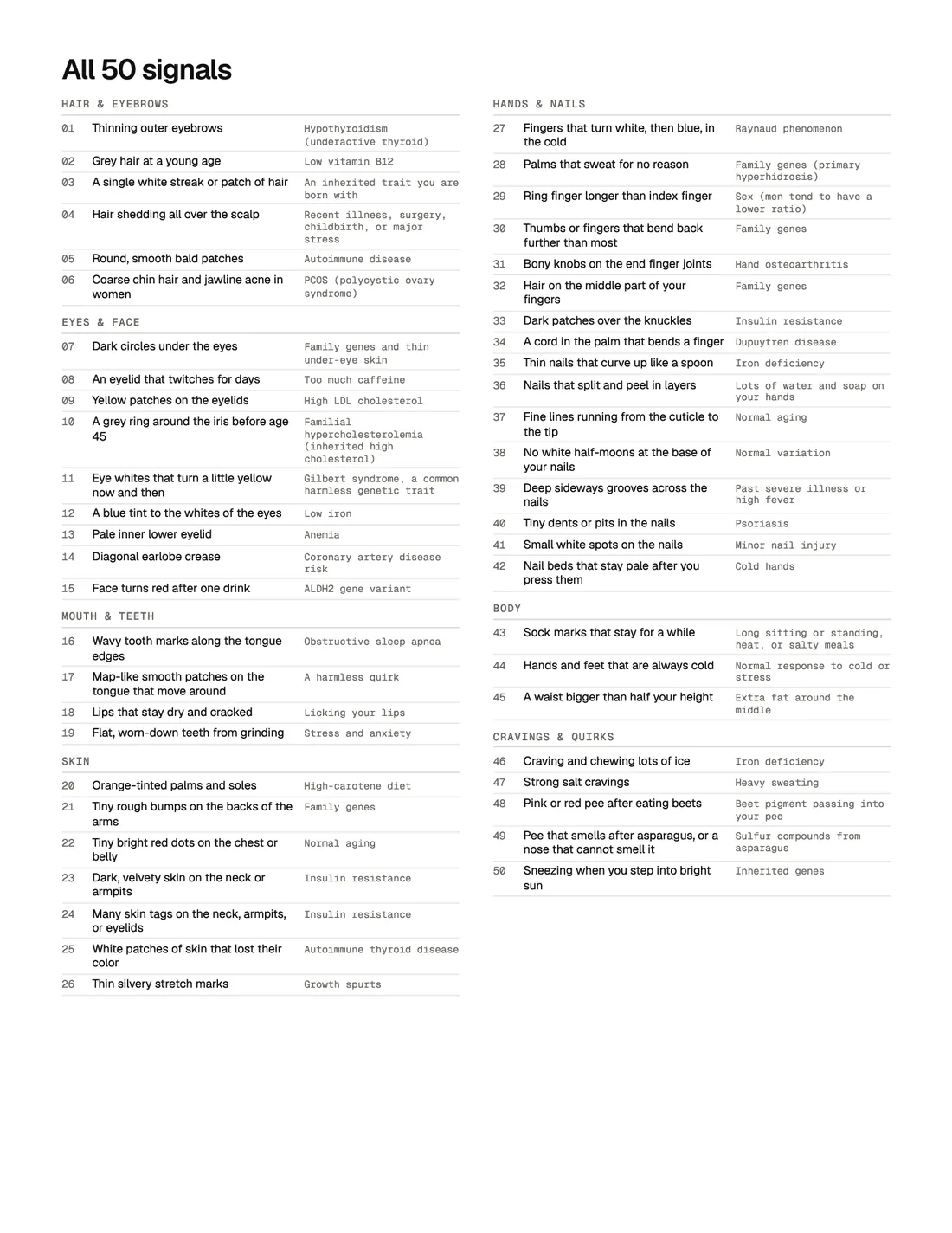
    All 50, on one spread

    Every signal with the thing it points to, so you can scan for the one you came here about.

  2. 02

    One signal, fully worked

    What to look for, why it happens, what else causes it, how strong the link is, and what to ask for.

  3. 03

    Most of them are harmless

    Plenty of entries end with nothing to test. The guide is as clear about those as it is about the rest.

Read this first

  1. Most of these are harmless. One quirk on its own is a fun fact. Two or three that point the same way, like low iron or a slow thyroid, are worth a blood test.
  2. Compare against your own normal. A change you notice matters more than a trait you have had your whole life.
  3. Every sign lists what to check. Bring the list to your next checkup. A photo of the change helps.
  4. A few are worth a visit. When a sign needs prompt care, you will see a box marked Get seen soon.

How serious is it

  • Just a quirkNothing to do. Fun to know.22
  • Mention at a checkupWorth a question or a blood test at your next routine visit.27
  • Book a visitGet it looked at soon, even if you feel fine.1

How strong is the evidence

  • Strong linkA textbook sign. Doctors look for it on purpose.20
  • Real linkStudies keep finding the connection, but plenty of other things cause it too.23
  • Early evidenceSmall studies point this way. Worth a test, not a conclusion.5
  • Mostly mythThe popular claim does not hold up. We tell you what does.2

All 50 signals

Hair & eyebrows

  1. 01Thinning outer eyebrowsHypothyroidism (underactive thyroid)
  2. 02Grey hair at a young ageLow vitamin B12
  3. 03A single white streak or patch of hairAn inherited trait you are born with
  4. 04Hair shedding all over the scalpRecent illness, surgery, childbirth, or major stress
  5. 05Round, smooth bald patchesAutoimmune disease
  6. 06Coarse chin hair and jawline acne in womenPCOS (polycystic ovary syndrome)

Eyes & face

  1. 07Dark circles under the eyesFamily genes and thin under-eye skin
  2. 08An eyelid that twitches for daysToo much caffeine
  3. 09Yellow patches on the eyelidsHigh LDL cholesterol
  4. 10A grey ring around the iris before age 45Familial hypercholesterolemia (inherited high cholesterol)
  5. 11Eye whites that turn a little yellow now and thenGilbert syndrome, a common harmless genetic trait
  6. 12A blue tint to the whites of the eyesLow iron
  7. 13Pale inner lower eyelidAnemia
  8. 14Diagonal earlobe creaseCoronary artery disease risk
  9. 15Face turns red after one drinkALDH2 gene variant

Mouth & teeth

  1. 16Wavy tooth marks along the tongue edgesObstructive sleep apnea
  2. 17Map-like smooth patches on the tongue that move aroundA harmless quirk
  3. 18Lips that stay dry and crackedLicking your lips
  4. 19Flat, worn-down teeth from grindingStress and anxiety

Skin

  1. 20Orange-tinted palms and solesHigh-carotene diet
  2. 21Tiny rough bumps on the backs of the armsFamily genes
  3. 22Tiny bright red dots on the chest or bellyNormal aging
  4. 23Dark, velvety skin on the neck or armpitsInsulin resistance
  5. 24Many skin tags on the neck, armpits, or eyelidsInsulin resistance
  6. 25White patches of skin that lost their colorAutoimmune thyroid disease
  7. 26Thin silvery stretch marksGrowth spurts

Hands & nails

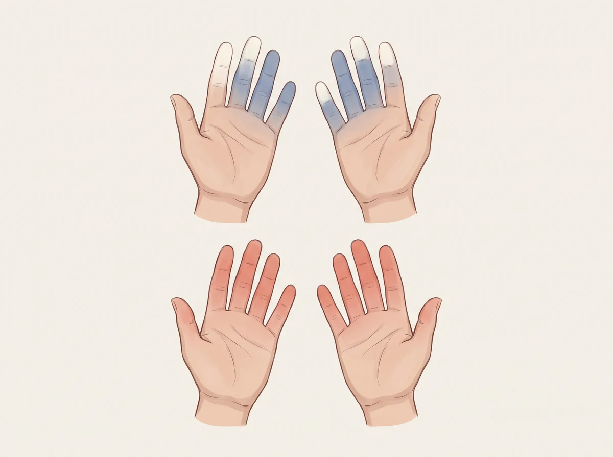
  1. 27Fingers that turn white, then blue, in the coldRaynaud phenomenon
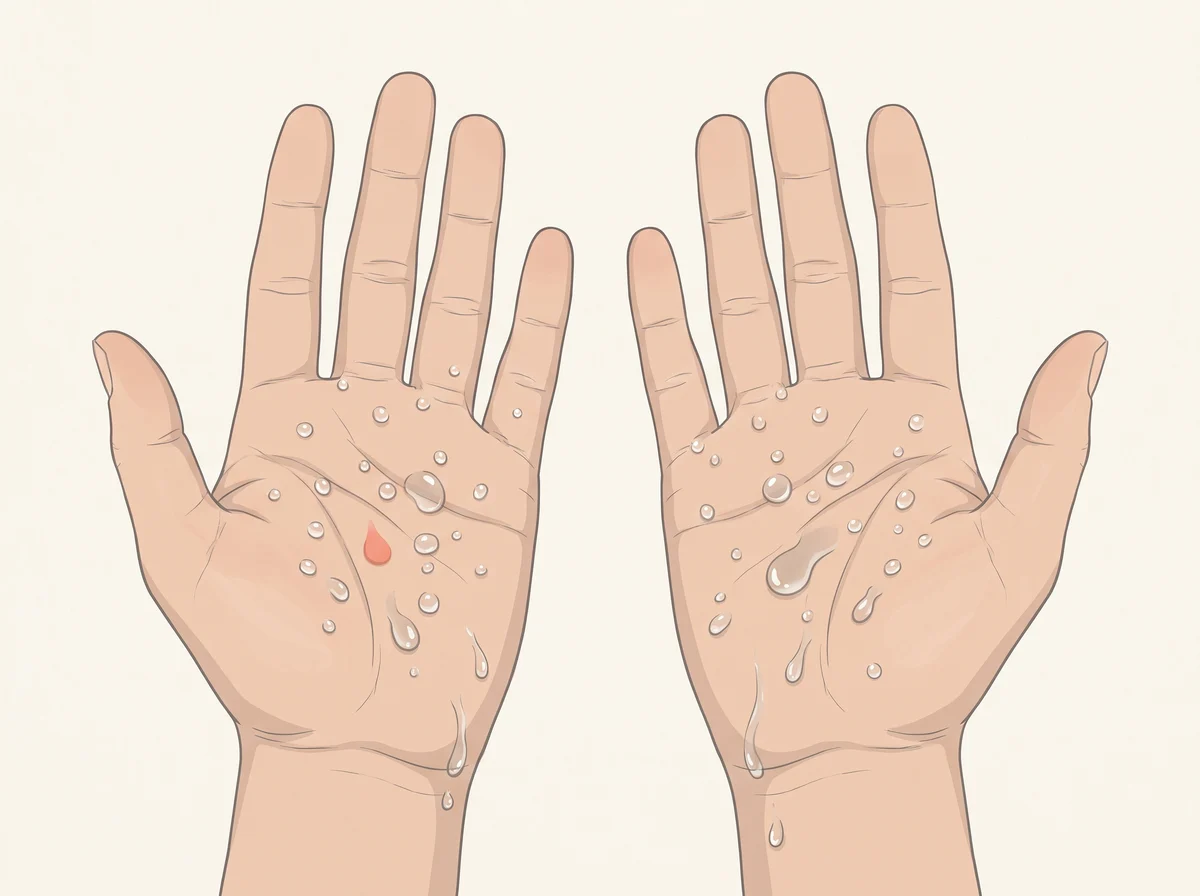
  2. 28Palms that sweat for no reasonFamily genes (primary hyperhidrosis)
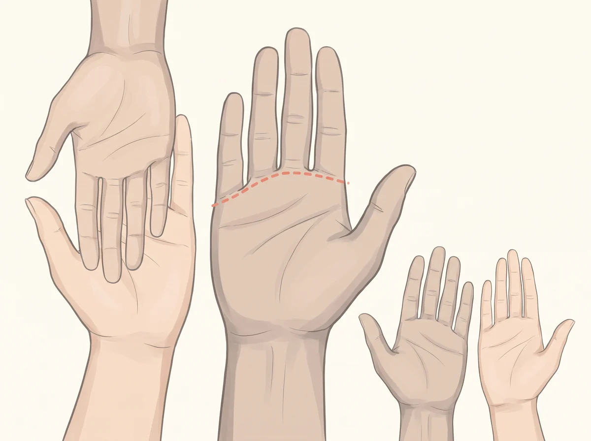
  3. 29Ring finger longer than index fingerSex (men tend to have a lower ratio)
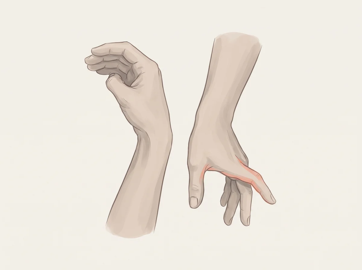
  4. 30Thumbs or fingers that bend back further than mostFamily genes
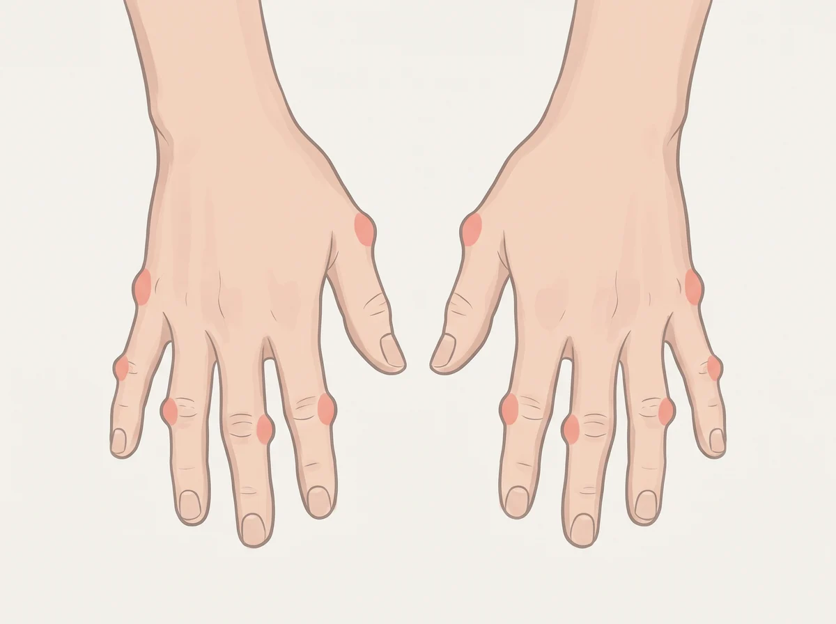
  5. 31Bony knobs on the end finger jointsHand osteoarthritis
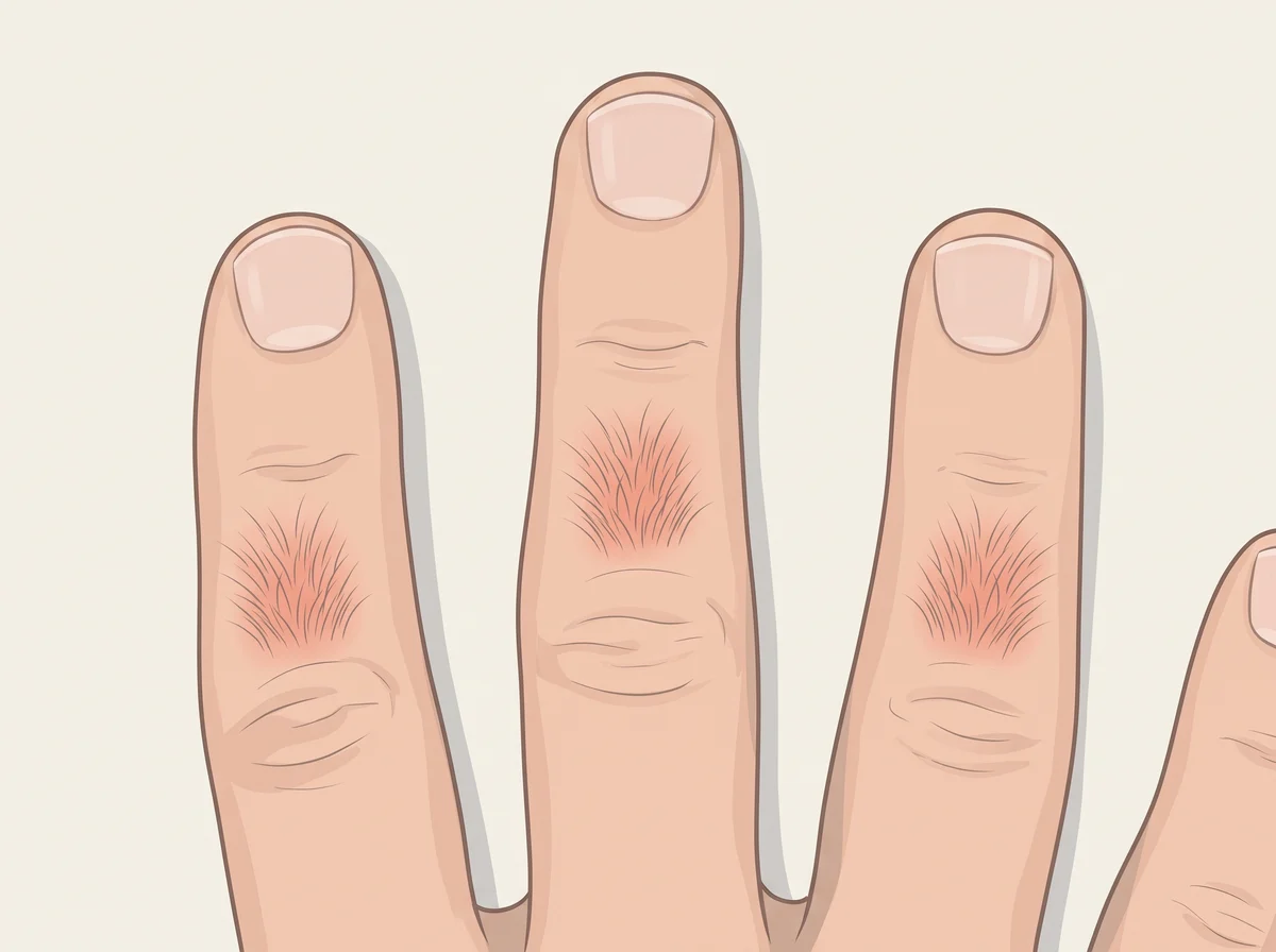
  6. 32Hair on the middle part of your fingersFamily genes
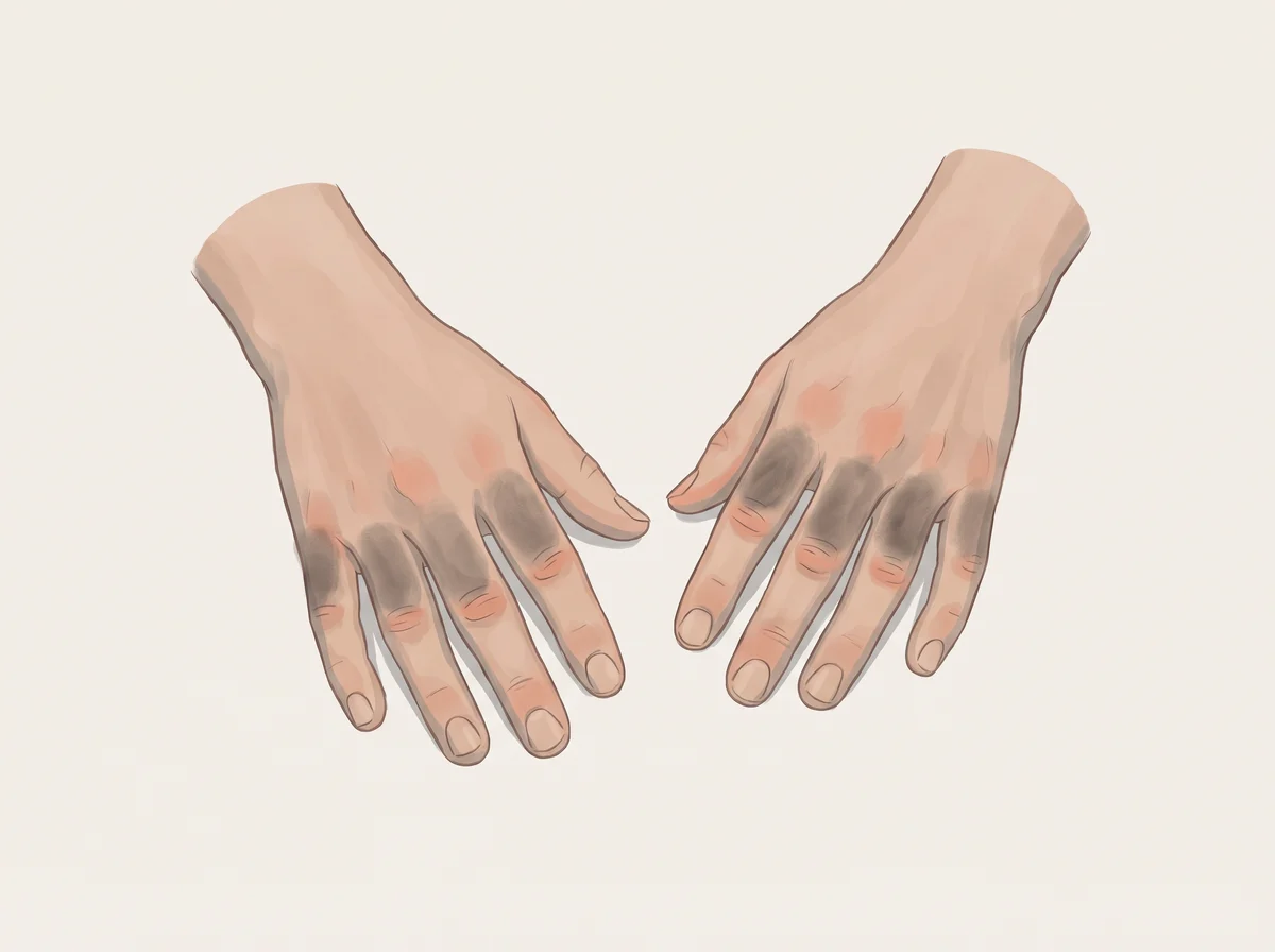
  7. 33Dark patches over the knucklesInsulin resistance
  8. 34A cord in the palm that bends a fingerDupuytren disease
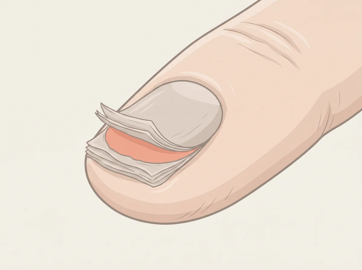
  9. 35Thin nails that curve up like a spoonIron deficiency
  10. 36Nails that split and peel in layersLots of water and soap on your hands
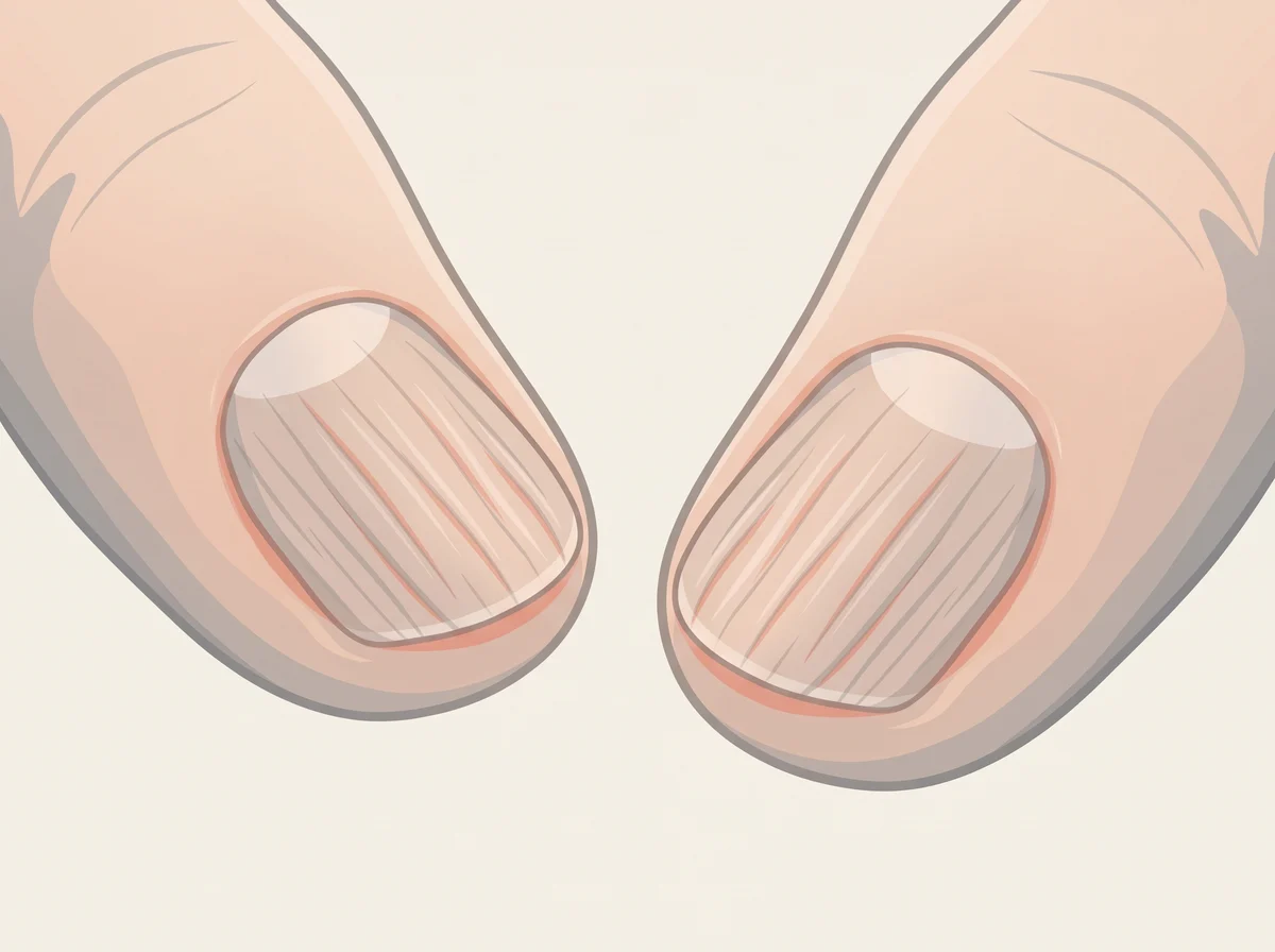
  11. 37Fine lines running from the cuticle to the tipNormal aging
  12. 38No white half-moons at the base of your nailsNormal variation
  13. 39Deep sideways grooves across the nailsPast severe illness or high fever
  14. 40Tiny dents or pits in the nailsPsoriasis
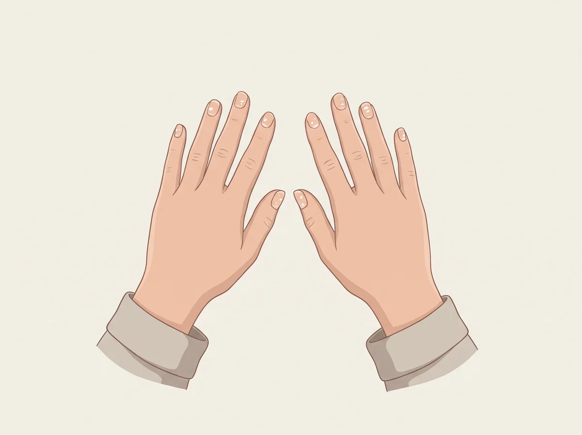
  15. 41Small white spots on the nailsMinor nail injury
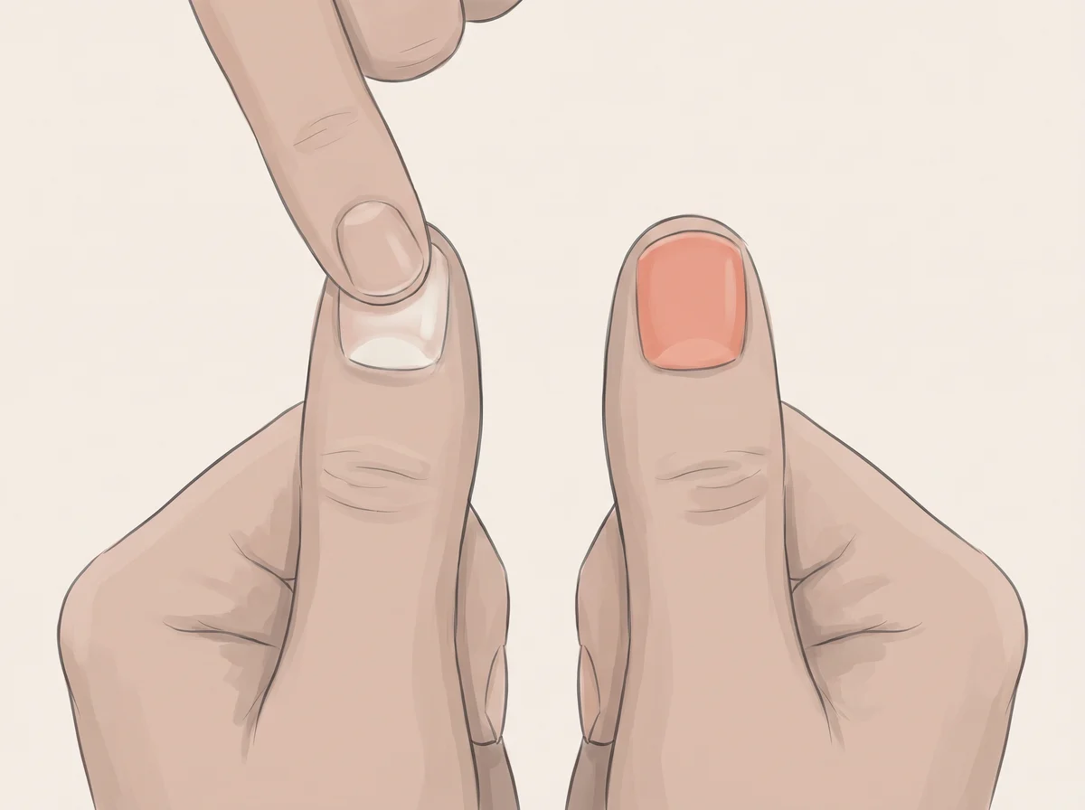
  16. 42Nail beds that stay pale after you press themCold hands

Body

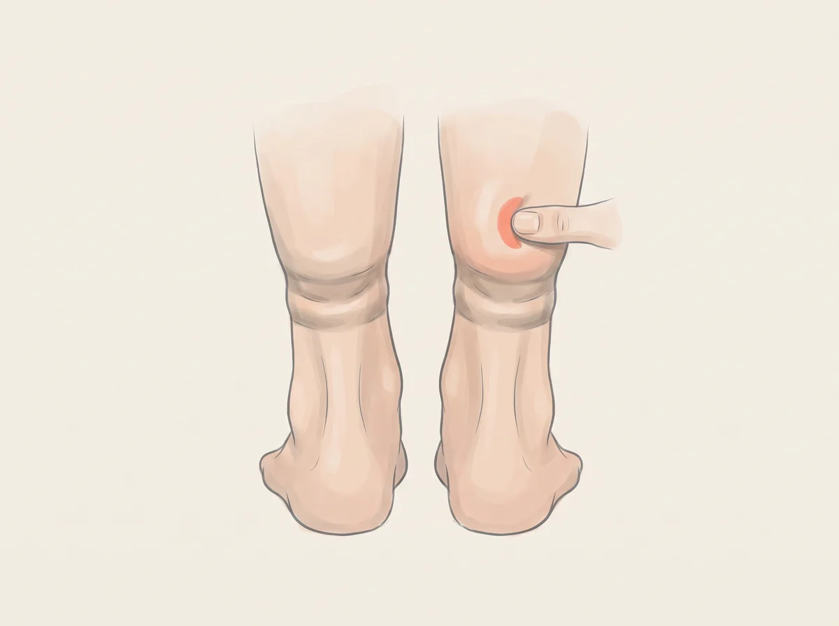
  1. 43Sock marks that stay for a whileLong sitting or standing, heat, or salty meals
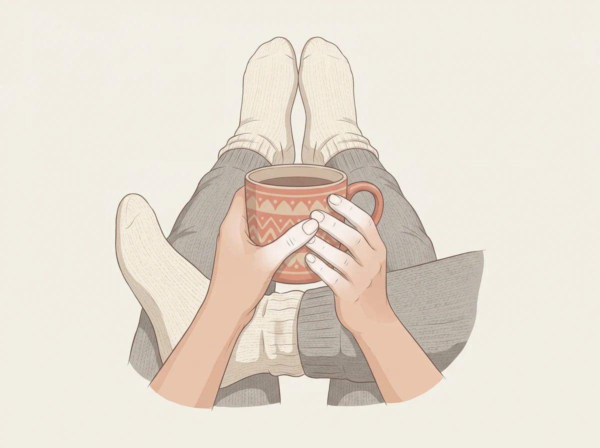
  2. 44Hands and feet that are always coldNormal response to cold or stress
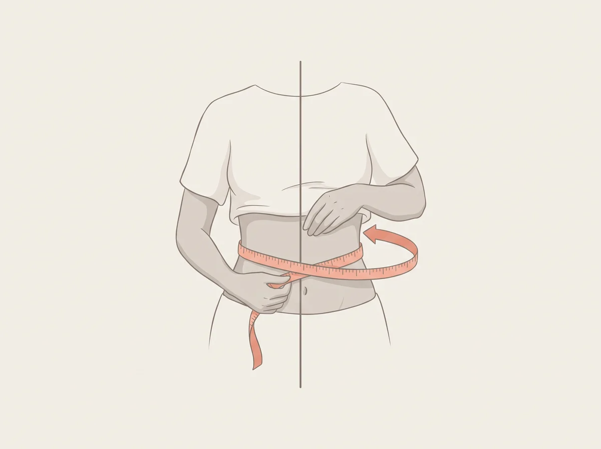
  3. 45A waist bigger than half your heightExtra fat around the middle

Cravings & quirks

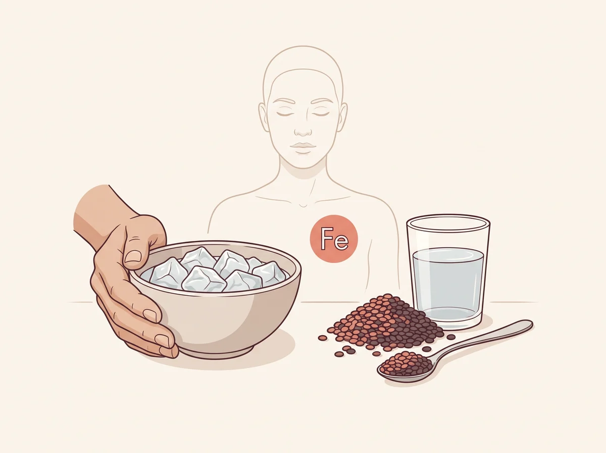
  1. 46Craving and chewing lots of iceIron deficiency
  2. 47Strong salt cravingsHeavy sweating
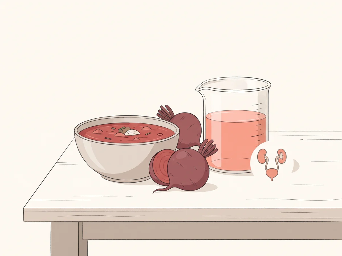
  3. 48Pink or red pee after eating beetsBeet pigment passing into your pee
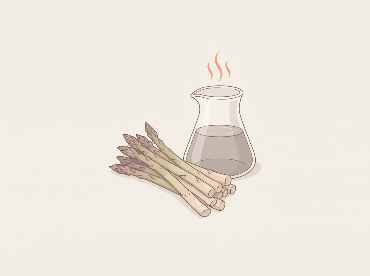
  4. 49Pee that smells after asparagus, or a nose that cannot smell itSulfur compounds from asparagus
  5. 50Sneezing when you step into bright sunInherited genes

Hair & eyebrows

01–06
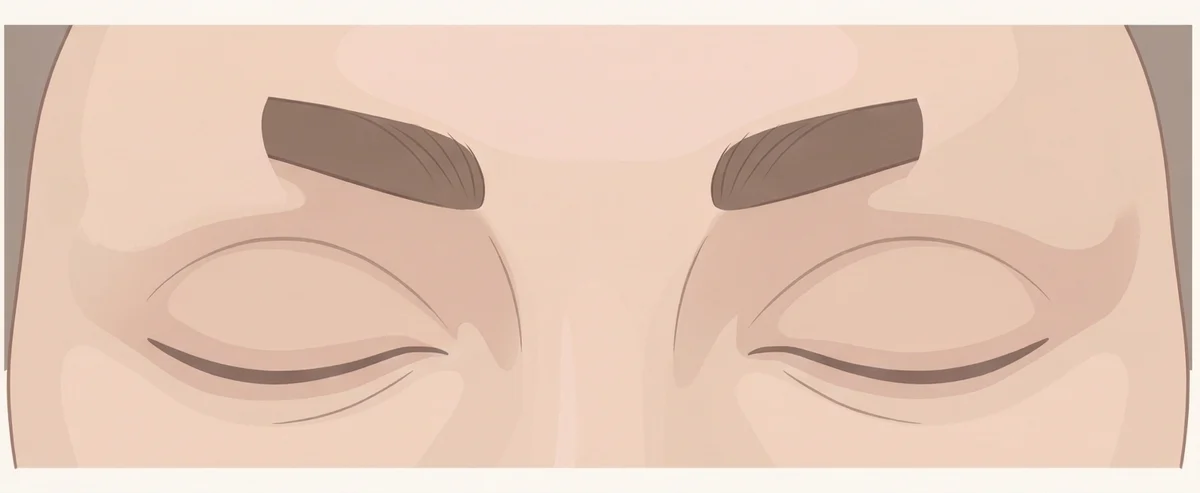
Illustration of thinning outer eyebrows. The outer third of the eyebrow, the part nearest the ear, gets thin or disappears on both sides.  The inner part of the brow stays fuller.
Illustration, not a photograph
01

Thinning outer eyebrows

Sign of Hertoghe (Queen Anne’s sign), a type of madarosis

The tail end of your eyebrows has a thyroid story to tell

Mention at a checkup

Linked to
  • Hypothyroidism (underactive thyroid)
  • Eczema around the eyes
  • Alopecia areata
What to look for
The outer third of the eyebrow, the part nearest the ear, gets thin or disappears on both sides. The inner part of the brow stays fuller.
Why it happens
Thyroid hormone keeps hair roots in their growing phase. When the thyroid makes too little hormone, the roots rest longer, and brow hair grows thin and coarse. A buildup of a sugar-protein gel in the skin may also play a part.
Other causes
Years of plucking or waxing, normal aging, and rubbing from eczema are far more common reasons. Alopecia areata and some infections can also thin the brows.
How strong is the link
Real link This is a classic textbook sign of hypothyroidism, but doctors describe it as uncommon, and no good study shows how often it appears. On its own it cannot diagnose a thyroid problem. It matters most when you also feel tired, cold, constipated, or gain weight without a clear reason.
What to check
  • TSH
  • Free T4
  • TPO antibodies (checks for Hashimoto thyroiditis)

Get seen soon Book a visit if thin brows come with heavy tiredness, feeling cold all the time, a puffy face, slow thinking, or a swelling in the front of your neck.

  1. Eyebrow loss and the Queen Anne sign in hypothyroidism · QJM: An International Journal of Medicine, 2023
  2. Madarosis (Eyebrow & Eyelash Hair Loss): Causes & Treatment · Cleveland Clinic, 2023
  3. Eyelash Loss: An Unusual Manifestation of Uncontrolled Hypothyroidism · Cureus (PubMed Central), 2024
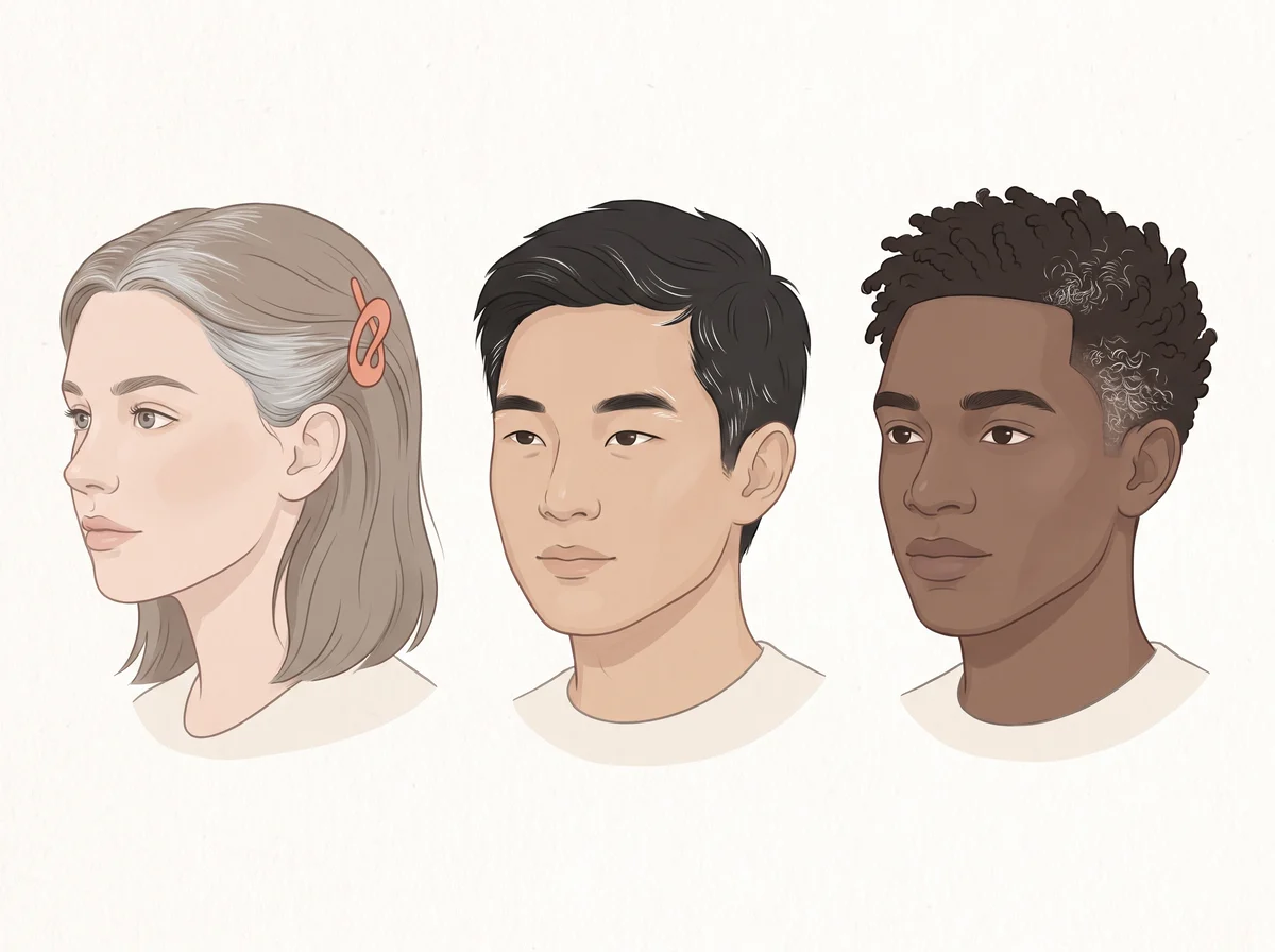
Illustration of grey hair at a young age. Many grey or white hairs before about age 20 in white people, 25 in Asian people, or 30 in Black people.  The grey often starts at the temples or the crown.
Illustration, not a photograph
02

Grey hair at a young age

Premature canities (premature hair greying)

Silver strands in your twenties are mostly a family trait

Just a quirk

Linked to
  • Low vitamin B12
  • Thyroid disease
  • Low vitamin D
  • Low ferritin or copper
What to look for
Many grey or white hairs before about age 20 in white people, 25 in Asian people, or 30 in Black people. The grey often starts at the temples or the crown.
Why it happens
Hair gets its color from pigment cells in the root. These cells need copper, B12, and other nutrients to make pigment, and wear and tear from everyday oxidative stress slows them down. When the cells tire out or run short, new hair grows in without color.
Other causes
Family history explains most early grey hair; twin studies suggest genes drive up to 90% of it. Smoking and vitiligo are also linked to early grey hair.
How strong is the link
Early evidence This is weak evidence. Small case-control studies from India and Iran, most with fewer than 75 patients, found lower B12, vitamin D, ferritin, or copper in young people with grey hair, but the results conflict: one study found no ferritin link and another found higher iron. The popular idea that grey hair means low copper rests on a few small studies.
What to check
  • Vitamin B12
  • TSH and free T4
  • 25-hydroxy vitamin D
  • Ferritin and complete blood count
  1. Premature hair graying: a multifaceted phenomenon · International Journal of Dermatology (PubMed Central), 2025
  2. Demographic Characteristics and Association of Serum Vitamin B12, Ferritin and Thyroid Function with Premature Canities in Indian Patients from an Urban Skin Clinic of North India · Indian Journal of Dermatology (PubMed Central), 2017
  3. Factors Associated with Premature Hair Graying in a Young Indian Population · International Journal of Trichology (PubMed Central), 2016
  4. Relationship between Trace Elements and Premature Hair Graying · International Journal of Trichology (PubMed Central), 2018
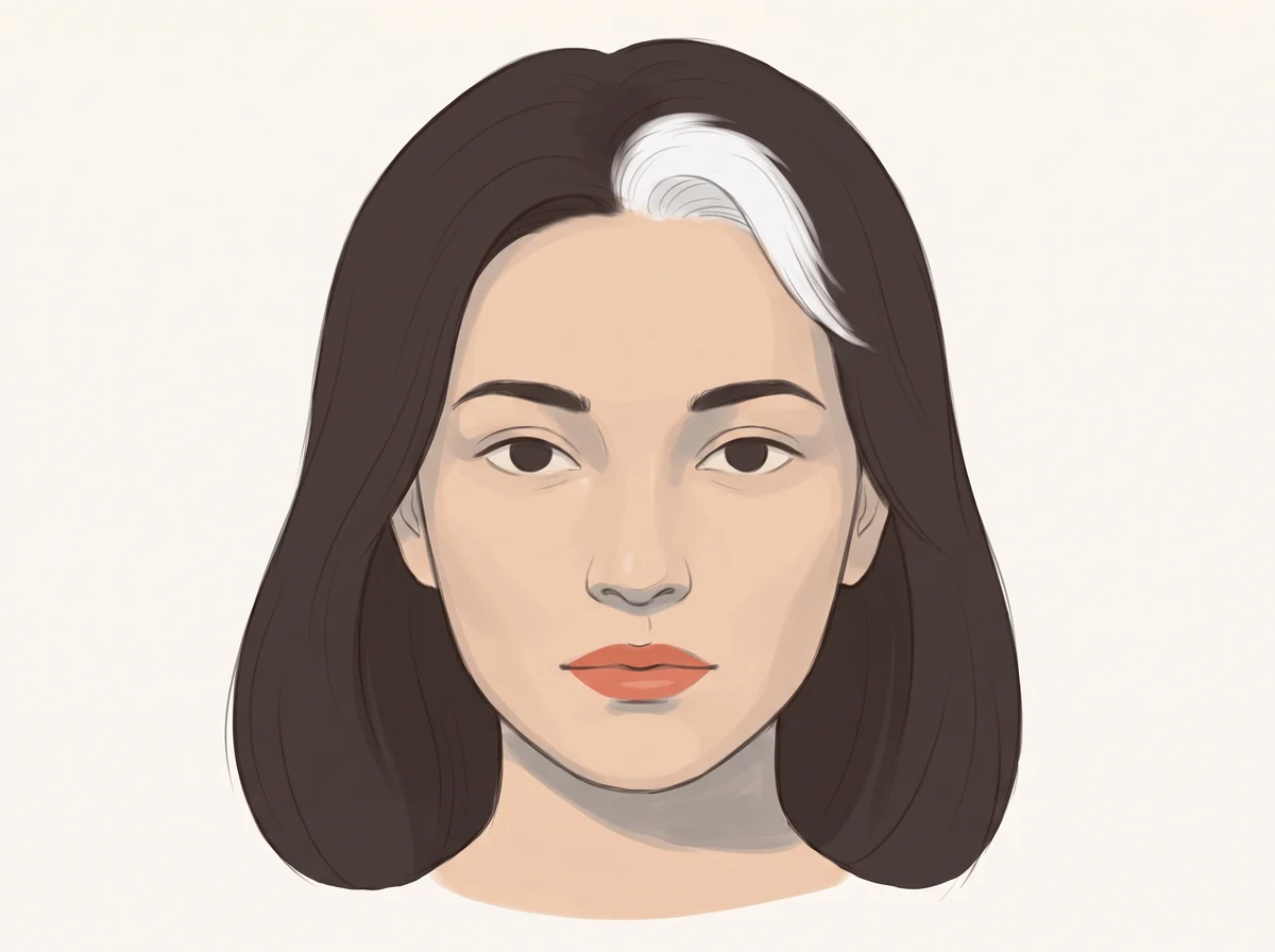
Illustration of a single white streak or patch of hair. One tidy patch of white or gray hair in hair that is otherwise dark.  It can be at the front hairline, elsewhere on the scalp, or in an eyebrow, eyelashes, or a beard.
Illustration, not a photograph
03

A single white streak or patch of hair

Poliosis (white forelock when it sits at the front hairline)

A skunk stripe of white hair is often just in your genes

Mention at a checkup

Linked to
  • An inherited trait you are born with
  • Vitiligo
  • Hair regrowing after alopecia areata
  • A mole under the patch (rare)
What to look for
One tidy patch of white or gray hair in hair that is otherwise dark. It can be at the front hairline, elsewhere on the scalp, or in an eyebrow, eyelashes, or a beard.
Why it happens
Hair gets its color from pigment cells at the root of each hair. In a white patch, a group of roots has little or no pigment, so those hairs grow in white. Some people are born with a white forelock that runs in the family and never spreads.
Other causes
Vitiligo, a condition where skin and hair lose color in patches, can turn the hair above a pale patch white. Hair that grows back after alopecia areata (patchy hair loss) often comes in white at first and may darken again over months.
How strong is the link
Real link Dermatology reviews link white hair patches to inherited traits, vitiligo, and regrowth after alopecia areata. They do not give a number for how often a lone streak is harmless, so a new one is worth a quick look from a doctor.
What to check
  • Had it since childhood and it never changed: nothing to test.
  • New in adulthood: ask a doctor to look at the scalp and skin under the patch.
  • Mention any pale skin patches or round bald spots somewhere else on your body.
  1. Poliosis circumscripta: overview and underlying causes · Journal of the American Academy of Dermatology, 2013
  2. Piebaldism · StatPearls, NCBI Bookshelf, 2023
  3. White hair in alopecia areata: Clinical forms and proposed physiopathologic mechanisms · Journal of the American Academy of Dermatology, 2023
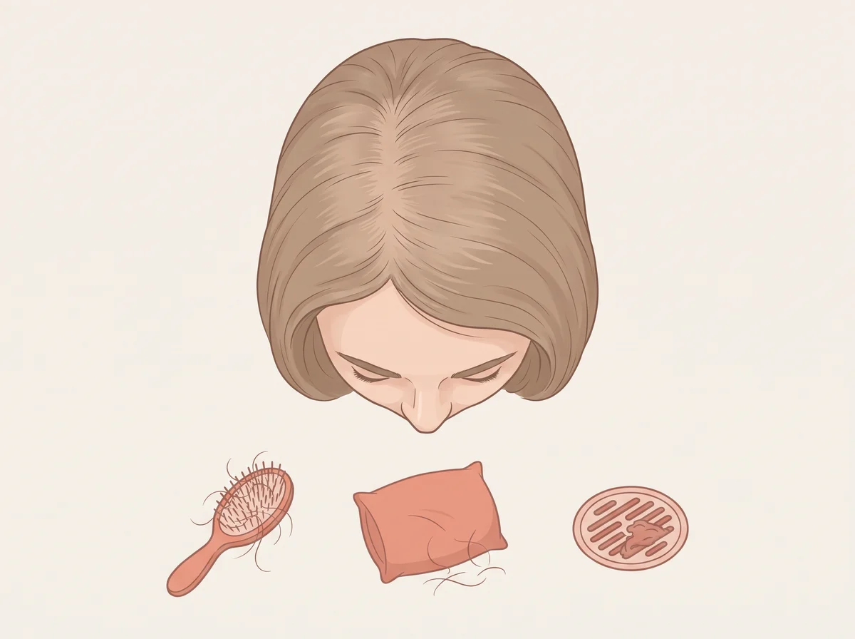
Illustration of hair shedding all over the scalp. More hair than usual in the shower drain, brush, or pillow, and a thinner ponytail or wider part.  The loss is spread over the whole scalp, with no bald spots.
Illustration, not a photograph
04

Hair shedding all over the scalp

Telogen effluvium

Extra hair in the drain often traces back about three months

Mention at a checkup

Linked to
  • Recent illness, surgery, childbirth, or major stress
  • Low ferritin (iron stores)
  • Thyroid disease
  • Crash dieting or low protein
What to look for
More hair than usual in the shower drain, brush, or pillow, and a thinner ponytail or wider part. The loss is spread over the whole scalp, with no bald spots.
Why it happens
A big event for the body, such as a high fever, surgery, childbirth, or a crash diet, sends many hair roots into their resting phase at the same time. About 3 months later, often 1 to 6 months, those resting hairs let go together. Low iron and an underactive thyroid can keep the shedding going.
Other causes
Shedding 50 to 100 hairs a day is normal. Stopping birth control pills and some medicines, such as beta-blockers and retinoids, can also trigger shedding.
How strong is the link
Real link The link to a recent shock such as illness or childbirth is well established, and the hair usually returns within 6 to 9 months once the trigger is gone. Low iron is common in people with this problem: one clinic study of 2,413 patients found low ferritin in 46.5%. But studies do not agree on whether low iron causes the shedding.
What to check
  • Ferritin, iron, and iron saturation
  • Complete blood count
  • TSH
  • A hair pull test by a dermatologist

Get seen soon See a doctor or dermatologist if shedding lasts more than 6 months, you spot bald patches, or it comes with heavy periods, weight loss, or deep tiredness.

  1. Telogen Effluvium · StatPearls, NCBI Bookshelf, 2024
  2. Hair shedding · American Academy of Dermatology
  3. Retrospective Review of 2851 Female Patients With Telogen Effluvium: A Single-Center Experience · Journal of Cosmetic Dermatology (PubMed Central), 2025
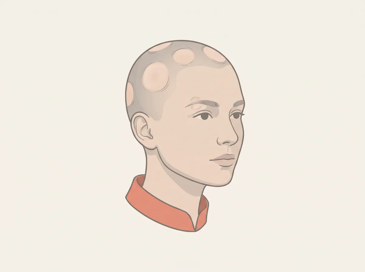
Illustration of round, smooth bald patches. One or more coin-sized, perfectly smooth bald spots on the scalp, beard, or eyebrows, often appearing within days.  The skin looks normal, with no scaling or scarring.
Illustration, not a photograph
05

Round, smooth bald patches

Alopecia areata

A coin-sized bald spot is your immune system pausing a few hairs

Book a visit

Linked to
  • Autoimmune disease
  • Thyroid autoimmunity (Hashimoto or Graves)
  • Vitiligo
  • Low vitamin D
What to look for
One or more coin-sized, perfectly smooth bald spots on the scalp, beard, or eyebrows, often appearing within days. The skin looks normal, with no scaling or scarring.
Why it happens
The immune system attacks hair roots by mistake, which is called an autoimmune reaction. The roots stay alive but take a break from making hair, so the hair can grow back. People with one autoimmune condition are more likely to have another.
Other causes
Fungal scalp infection (ringworm), hair pulling, and tight hairstyles can also cause patches, but these usually leave broken hairs, scaling, or redness.
How strong is the link
Strong link Alopecia areata is an autoimmune disease, and its link to thyroid antibodies is well documented: a review of 17 studies found higher rates of TPO and thyroglobulin antibodies, though not a clear rise in diagnosed thyroid disease. The vitamin D link is weaker: people with alopecia areata have lower average levels, but no one has shown that low vitamin D causes it.
What to check
  • TSH
  • TPO and thyroglobulin antibodies
  • 25-hydroxy vitamin D
  • A skin exam for vitiligo patches

Get seen soon See a dermatologist soon if patches spread quickly, join together, or you lose eyebrows, eyelashes, or body hair. Early treatment works best.

  1. Alopecia areata: Causes · American Academy of Dermatology
  2. Chronological association between alopecia areata and autoimmune thyroid diseases: A systematic review and meta-analysis · The Journal of Dermatology (PubMed), 2019
  3. Increased prevalence of vitamin D deficiency in patients with alopecia areata: a systematic review and meta-analysis · Journal of the European Academy of Dermatology and Venereology (PubMed), 2018
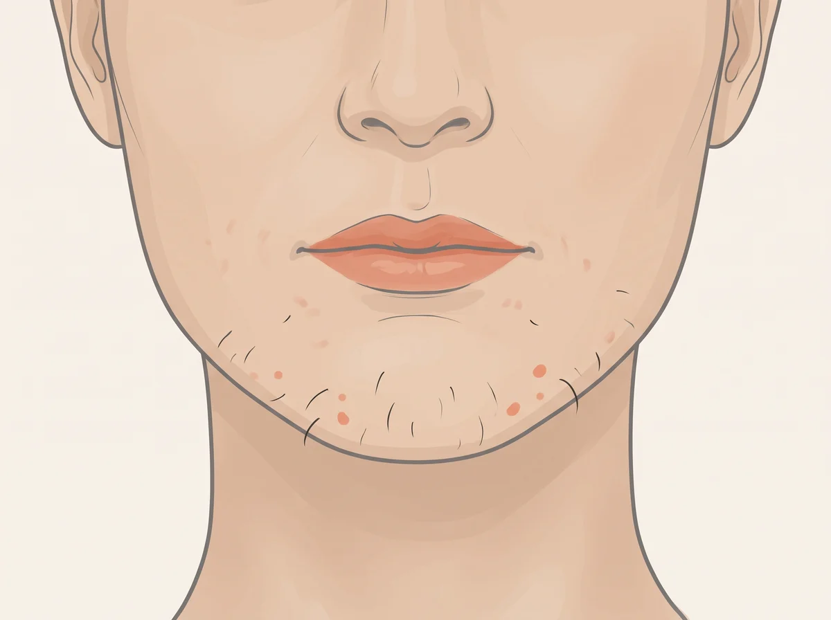
Illustration of coarse chin hair and jawline acne in women. Thick, dark hairs on the chin, upper lip, jawline, chest, or belly, in the pattern where men grow hair.  Some women also get deep, stubborn pimples along the jaw and chin.
Illustration, not a photograph
06

Coarse chin hair and jawline acne in women

Hirsutism and hyperandrogenism

Dark chin hairs in women are a common hormone clue, often PCOS

Mention at a checkup

Linked to
  • PCOS (polycystic ovary syndrome)
  • High androgens (male-type hormones)
  • Insulin resistance
  • Adrenal gland disorders
What to look for
Thick, dark hairs on the chin, upper lip, jawline, chest, or belly, in the pattern where men grow hair. Some women also get deep, stubborn pimples along the jaw and chin.
Why it happens
Androgens are hormones like testosterone that women make in small amounts. In PCOS the ovaries make more, often because high insulin levels push them to. Extra androgen turns fine, pale body hair into thick, dark hair and makes the skin oilier.
Other causes
Some women with normal hormones have extra hair from family background or ethnicity, which doctors call idiopathic hirsutism. Medicines such as minoxidil, phenytoin, and anabolic steroids can also cause it.
How strong is the link
Strong link The hair is the strong part: about 1 in 10 women has hirsutism, and PCOS causes roughly 3 in 4 cases. Acne alone is a weaker clue. Experts disagree on whether acne without extra hair means high androgens, though one study of 207 women with acne found 72% had an androgen problem.
What to check
  • Total and free testosterone
  • DHEA-S
  • 17-hydroxyprogesterone
  • Fasting glucose, HbA1c, and fasting insulin; plus prolactin, TSH, and a pelvic ultrasound if your doctor suggests

Get seen soon Book a visit soon if hair growth starts suddenly or gets much heavier over a few months, or comes with a deeper voice, balding at the temples, or an enlarged clitoris. A rare hormone-making tumor can cause this faster pattern.

  1. Hirsutism · StatPearls, NCBI Bookshelf, 2023
  2. Epidemiology, diagnosis and management of hirsutism: a consensus statement by the Androgen Excess and Polycystic Ovary Syndrome Society · Human Reproduction Update (PubMed), 2012
  3. Hirsutism and Polycystic Ovary Syndrome (PCOS) · American Society for Reproductive Medicine (ReproductiveFacts.org), 2016
  4. Is acne a sign of androgen excess disorder or not? · European Journal of Obstetrics, Gynecology, and Reproductive Biology (PubMed), 2017

Eyes & face

07–15
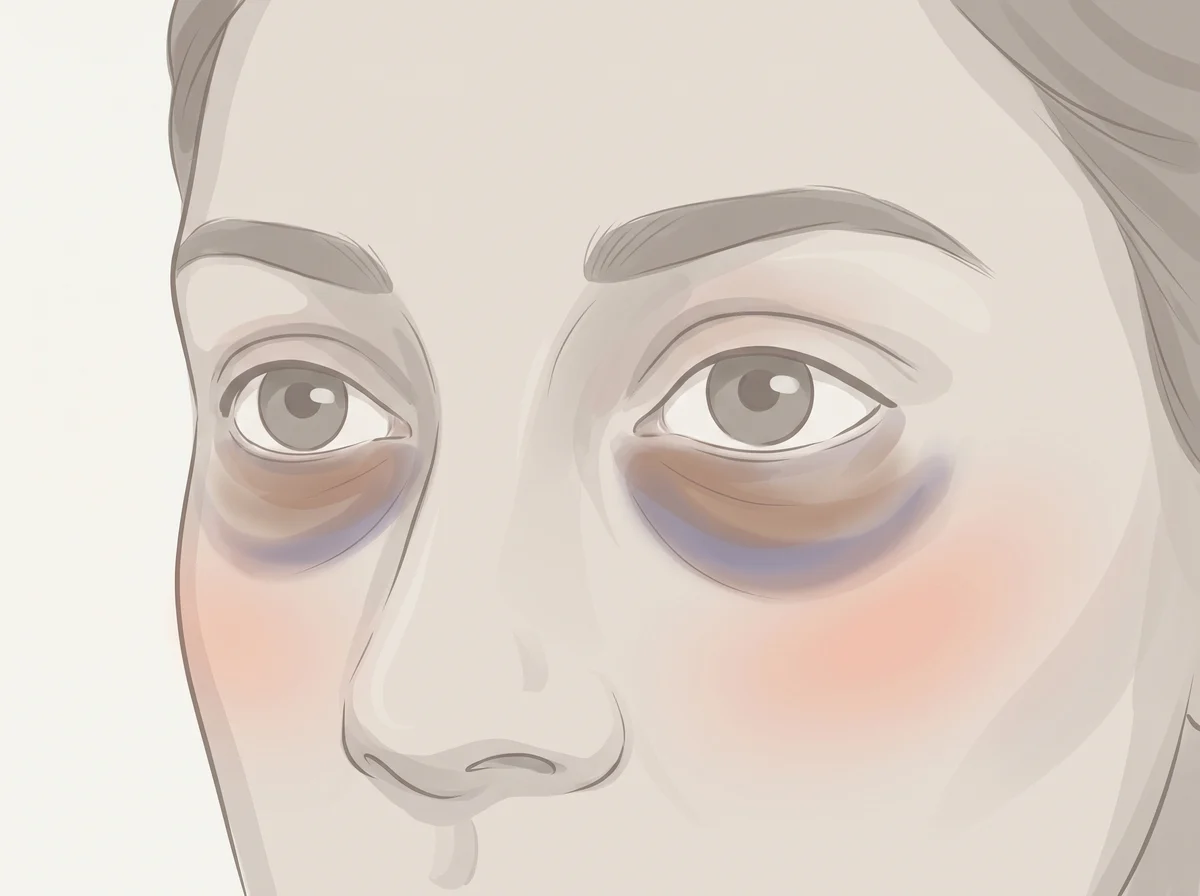
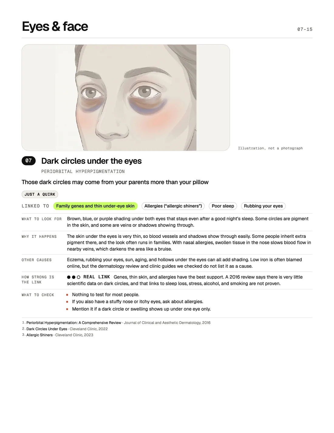
Illustration of dark circles under the eyes. Brown, blue, or purple shading under both eyes that stays even after a good night’s sleep.  Some circles are pigment in the skin, and some are veins or shadows showing through.
Illustration, not a photograph
07

Dark circles under the eyes

Periorbital hyperpigmentation

Those dark circles may come from your parents more than your pillow

Just a quirk

Linked to
  • Family genes and thin under-eye skin
  • Allergies (“allergic shiners”)
  • Poor sleep
  • Rubbing your eyes
What to look for
Brown, blue, or purple shading under both eyes that stays even after a good night’s sleep. Some circles are pigment in the skin, and some are veins or shadows showing through.
Why it happens
The skin under the eyes is very thin, so blood vessels and shadows show through easily. Some people inherit extra pigment there, and the look often runs in families. With nasal allergies, swollen tissue in the nose slows blood flow in nearby veins, which darkens the area like a bruise.
Other causes
Eczema, rubbing your eyes, sun, aging, and hollows under the eyes can all add shading. Low iron is often blamed online, but the dermatology review and clinic guides we checked do not list it as a cause.
How strong is the link
Real link Genes, thin skin, and allergies have the best support. A 2016 review says there is very little scientific data on dark circles, and that links to sleep loss, stress, alcohol, and smoking are not proven.
What to check
  • Nothing to test for most people.
  • If you also have a stuffy nose or itchy eyes, ask about allergies.
  • Mention it if a dark circle or swelling shows up under one eye only.
  1. Periorbital Hyperpigmentation: A Comprehensive Review · Journal of Clinical and Aesthetic Dermatology, 2016
  2. Dark Circles Under Eyes · Cleveland Clinic, 2022
  3. Allergic Shiners · Cleveland Clinic, 2023
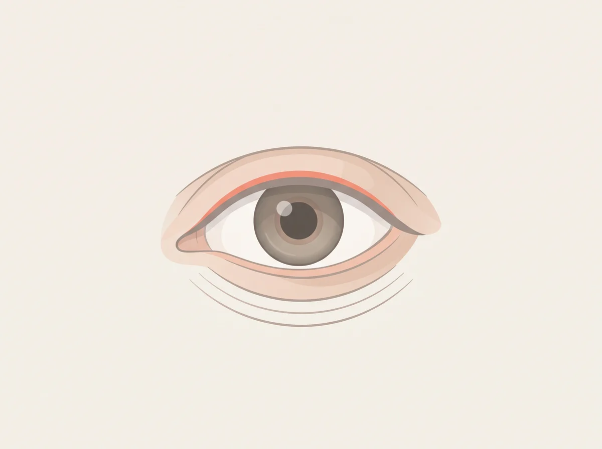
Illustration of an eyelid that twitches for days. A fine, rippling flicker, most often in the lower eyelid of one eye.  Other people usually cannot see it, and it comes and goes for seconds to hours.
Illustration, not a photograph
08

An eyelid that twitches for days

Eyelid myokymia

That eyelid flutter is your body asking for less coffee and more sleep

Just a quirk

Linked to
  • Too much caffeine
  • Stress
  • Short sleep and fatigue
  • Dry eyes
What to look for
A fine, rippling flicker, most often in the lower eyelid of one eye. Other people usually cannot see it, and it comes and goes for seconds to hours.
Why it happens
The ring of muscle around the eye starts firing on its own in small, repeated bursts. Doctors do not know the exact cause, but it shows up with stress, fatigue, and caffeine. It is so common during exam season that it has been nicknamed “the medical students’ disease.”
Other causes
Dry eyes, nicotine, alcohol, and hard exercise are also listed as triggers.
How strong is the link
Real link The trigger list comes from clinical experience more than large studies. The good news is well established: the twitch usually fades on its own and rarely points to any nerve disease.
What to check
  • Nothing to test. Cut back on caffeine and get more sleep for a week or two.
  • Mention it if the twitch lasts more than a few weeks or spreads to other parts of your face.

Get seen soon Get it checked soon if your eyelid closes all the way with each twitch, the lid droops, or other face muscles start to twitch.

  1. Eyelid Myokymia · StatPearls, NCBI Bookshelf, 2023
  2. Eyelid Myokymia · Cleveland Clinic, 2025
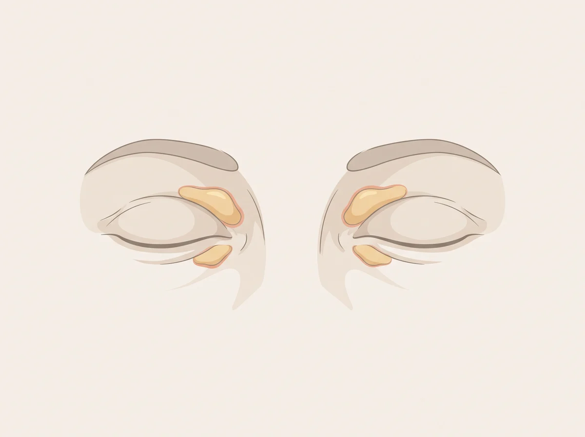
Illustration of yellow patches on the eyelids. Soft, flat or slightly raised yellow patches on the upper or lower eyelids, usually near the nose.  They are painless and grow slowly.
Illustration, not a photograph
09

Yellow patches on the eyelids

Xanthelasma palpebrarum

Little yellow eyelid patches are a nudge to check your cholesterol

Mention at a checkup

Linked to
  • High LDL cholesterol
  • Inherited lipid disorders
  • Higher heart attack risk
  • Hypothyroidism or diabetes
What to look for
Soft, flat or slightly raised yellow patches on the upper or lower eyelids, usually near the nose. They are painless and grow slowly.
Why it happens
The patches are small clusters of cells filled with cholesterol, sitting in the eyelid skin. High levels of LDL, the cholesterol linked to artery buildup, make these deposits more likely. The patches show that fat is settling in tissue, which is why doctors check the blood too.
Other causes
About half of people with these patches have normal cholesterol. Liver disease that blocks bile flow and kidney problems that leak protein can also raise blood fats and cause them.
How strong is the link
Real link About 50% of adults with xanthelasma have abnormal blood fats. A Danish study that followed 12,745 people for over 30 years found those with the patches had about 48% higher risk of heart attack, even after accounting for cholesterol. The patches themselves are harmless; the value is as a prompt to test your blood fats.
What to check
  • Fasting lipid panel (LDL, HDL, triglycerides)
  • Lipoprotein(a) and ApoB
  • TSH
  • Fasting glucose or HbA1c, and liver function tests

Get seen soon Get a cholesterol test soon if you are under 40 or close family had early heart attacks. See a doctor promptly for chest pain with exercise.

  1. Xanthelasma Palpebrarum · StatPearls, NCBI Bookshelf, 2026
  2. Xanthelasmata, arcus corneae, and ischaemic vascular disease and death in general population: prospective cohort study · BMJ (PubMed), 2011
  3. Serum lipids and risk of atherosclerosis in xanthelasma palpebrarum: A systematic review and meta-analysis · Journal of the American Academy of Dermatology (PubMed), 2020
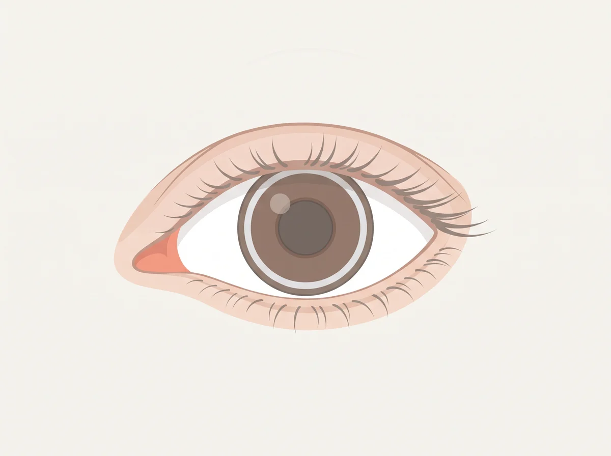
Illustration of a grey ring around the iris before age 45. A grey, white, or bluish ring at the outer edge of the colored part of the eye, often with a thin clear gap next to the white.  It usually starts at the top and bottom and fills in to a full circle.
Illustration, not a photograph
10

A grey ring around the iris before age 45

Corneal arcus (arcus juvenilis when young)

A grey ring around the iris is normal at 80 and a clue at 30

Mention at a checkup

Linked to
  • Familial hypercholesterolemia (inherited high cholesterol)
  • High LDL cholesterol
  • Early heart disease risk
What to look for
A grey, white, or bluish ring at the outer edge of the colored part of the eye, often with a thin clear gap next to the white. It usually starts at the top and bottom and fills in to a full circle.
Why it happens
Cholesterol and other fats seep from blood vessels at the edge of the cornea, the clear front window of the eye, and settle there. In young people this usually takes very high LDL levels for many years. Familial hypercholesterolemia is a gene change that keeps LDL high from birth.
Other causes
In older adults this ring is a normal part of aging; almost everyone over 80 has it, and it does not need testing past about age 50. It does not affect vision.
How strong is the link
Strong link Doctors use this ring in the standard scoring tool for inherited high cholesterol (the Dutch Lipid Clinic Network score), where a ring before age 45 adds 4 points. Many people with the inherited condition never get the ring, so a clear eye does not rule it out.
What to check
  • Fasting lipid panel with LDL cholesterol
  • Lipoprotein(a)
  • Family history of early heart attacks or high cholesterol
  • Genetic testing for familial hypercholesterolemia if LDL is very high

Get seen soon Ask for a cholesterol test soon if you are under 45 and see this ring, especially if a parent or sibling had a heart attack before 55 (men) or 60 (women).

  1. Arcus Senilis · StatPearls, NCBI Bookshelf, 2023
  2. Correlating corneal arcus with atherosclerosis in familial hypercholesterolemia · Lipids in Health and Disease (PubMed Central), 2008
  3. Dutch Lipid Clinic Network Score (DLCNS) for FH · FH Australasia Network (Australian Atherosclerosis Society)
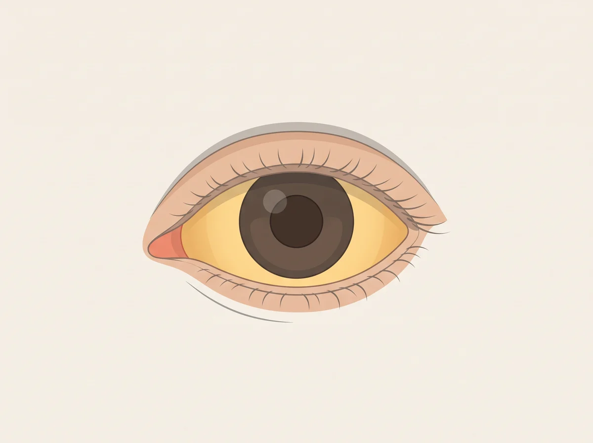
Illustration of eye whites that turn a little yellow now and then. The whites of both eyes take on a faint yellow tint for a few days, then clear up again.  It tends to show up when you skip meals, get sick, feel run down, sleep poorly, drink too little, exercise hard, or have your period.
Illustration, not a photograph
11

Eye whites that turn a little yellow now and then

Gilbert syndrome (mild scleral icterus)

Eyes that go a bit yellow on hungry, tired days are often Gilbert’s

Mention at a checkup

Linked to
  • Gilbert syndrome, a common harmless genetic trait
  • Liver problems such as hepatitis
  • Blocked bile duct
  • Fast breakdown of red blood cells (hemolysis)
What to look for
The whites of both eyes take on a faint yellow tint for a few days, then clear up again. It tends to show up when you skip meals, get sick, feel run down, sleep poorly, drink too little, exercise hard, or have your period. Daylight makes it easiest to see.
Why it happens
Bilirubin is a yellow pigment made when old red blood cells break down, and the liver clears it into bile. In Gilbert syndrome, a small change in the UGT1A1 gene makes the liver slower at this job, so a little extra bilirubin stays in the blood. Fasting or illness adds a bit more, and when levels climb high enough, the eye whites pick up the color.
Other causes
Yellow eyes can also come from liver problems such as hepatitis, a blocked bile duct, or red blood cells breaking down too fast. These look the same at first, so the first time you notice yellow eyes, a blood test sorts out which one it is. After a doctor confirms Gilbert syndrome, a mild yellow tint during a fast or a cold is expected.
How strong is the link
Strong link Gilbert syndrome is common: MedlinePlus puts it at 3 to 7 percent of Americans, and StatPearls gives 2 to 20 percent depending on ancestry. StatPearls and the NHS say it does not damage the liver and needs no treatment. The eye color alone cannot tell Gilbert syndrome apart from liver disease, which is why the first episode needs blood tests.
What to check
  • Total and direct bilirubin
  • Liver panel (ALT, AST, alkaline phosphatase, albumin)
  • Complete blood count with reticulocyte count, LDH, and haptoglobin
  • Abdominal ultrasound, if your doctor suggests it

Get seen soon If you have never been told you have Gilbert syndrome, see a doctor within a day or two for new yellow eyes. Go the same day if yellow eyes come with fever, belly pain, confusion, or pale stools with dark urine.

  1. Gilbert Syndrome · StatPearls, NCBI Bookshelf, 2024
  2. Gilbert syndrome · MedlinePlus Genetics, U.S. National Library of Medicine
  3. Gilbert’s syndrome · NHS, 2025
  4. Hyperbilirubinemia · StatPearls, NCBI Bookshelf, 2023
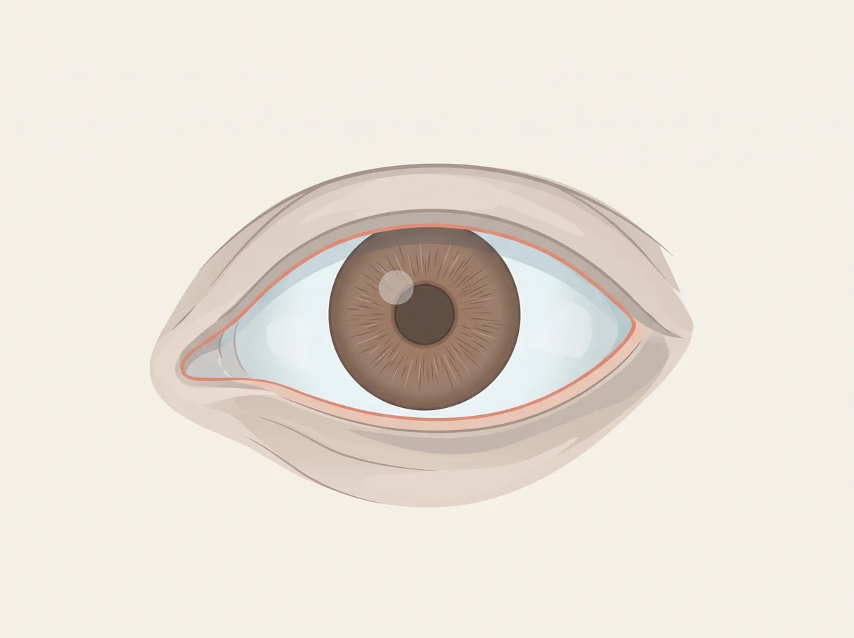
Illustration of a blue tint to the whites of the eyes. In daylight, the white part of the eye has a faint blue or gray tint.  It is easiest to judge side by side with someone else’s eyes in the same light.
Illustration, not a photograph
12

A blue tint to the whites of the eyes

Blue sclerae

Bluish eye whites can be a quiet hint of low iron

Mention at a checkup

Linked to
  • Low iron
  • Thin eye whites
  • Rare inherited collagen conditions
  • Some medicines
What to look for
In daylight, the white part of the eye has a faint blue or gray tint. It is easiest to judge side by side with someone else’s eyes in the same light.
Why it happens
The eye white (sclera) is made mostly of collagen. The body needs iron to build that collagen, so low iron may leave the white layer thinner. The dark layer underneath then shows through as blue.
Other causes
Rare inherited conditions that weaken collagen, such as osteogenesis imperfecta, make the eye whites clearly blue. The antibiotic minocycline and long-term steroid use have also been reported to cause it.
How strong is the link
Early evidence In a 1986 study of 169 hospital patients, 87% of those with iron deficiency anemia had blue eye whites, compared with about 5% of those without anemia. A 2019 pilot study of 67 patients found doctors often disagreed about who had the tint, so treat it as a hint and let a blood test decide.
What to check
  • Complete blood count (CBC)
  • Ferritin, which shows how much iron your body has stored
  1. Blue sclerae: a common sign of iron deficiency? · The Lancet, 1986
  2. Computed and Subjective Blue Scleral Color Analysis as a Diagnostic Tool for Iron Deficiency: A Pilot Study · Journal of Clinical Medicine, 2019
  3. Blue sclera: An overlooked finding of iron deficiency · Cleveland Clinic Journal of Medicine, 2022
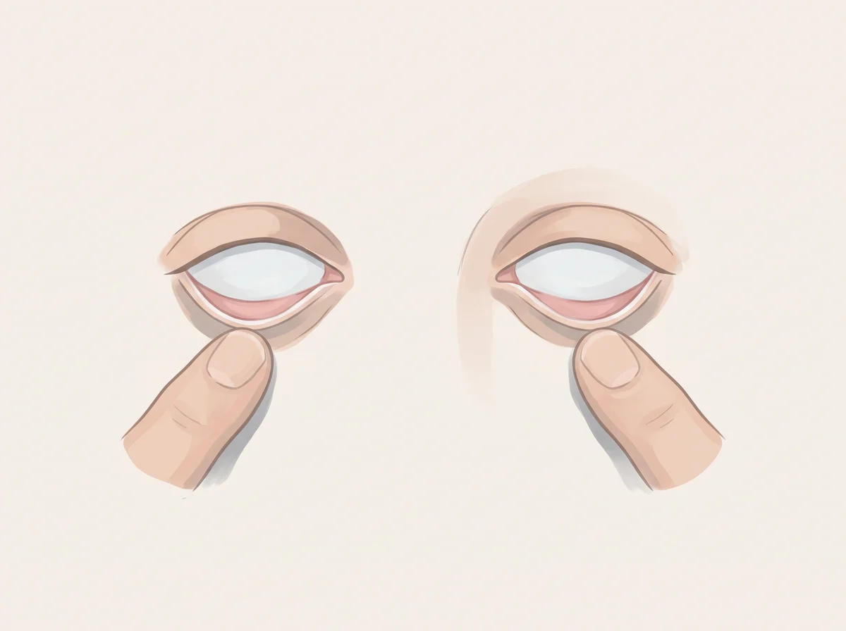
Illustration of pale inner lower eyelid. Gently pull down your lower eyelid in good light.  The inner lining is normally bright pink or red.
Illustration, not a photograph
13

Pale inner lower eyelid

Conjunctival pallor

Pull down your lower eyelid for a quick peek at your blood color

Mention at a checkup

Linked to
  • Anemia
  • Iron deficiency
  • Low vitamin B12 or folate
  • Blood loss, such as heavy periods
What to look for
Gently pull down your lower eyelid in good light. The inner lining is normally bright pink or red. With anemia it looks pale pink or almost white, most of all along the front rim.
Why it happens
Red blood cells carry hemoglobin, the red protein that moves oxygen. The inner eyelid has thin skin over many tiny blood vessels, so it shows blood color well. When hemoglobin drops, the lining loses its red color.
Other causes
Cold, poor lighting, and differences in how people judge color can make a normal eyelid look pale. Eye irritation or allergies can make a pale lid look red and hide anemia.
How strong is the link
Real link Pallor is a real sign of anemia, but a weak test: in one study it caught under 1 in 5 people with anemia, and doctors looking at the same eyelid often disagree. It works best for severe anemia, and a normal-looking eyelid does not rule anemia out.
What to check
  • Complete blood count (hemoglobin and red cell size)
  • Ferritin and iron studies
  • Vitamin B12 and folate
  • Stool test for hidden blood if iron is low without a clear cause

Get seen soon See a doctor soon if pale eyelids come with shortness of breath, a racing heart, dizziness, black or bloody stools, or very heavy periods.

  1. Accuracy and Reliability of Pallor for Detecting Anaemia: A Hospital-Based Diagnostic Accuracy Study · PLOS ONE (PubMed Central), 2010
  2. Accuracy of anemia diagnosis by physical examination · São Paulo Medical Journal (PubMed Central), 2007
  3. Iron deficiency anaemia · NHS, 2024
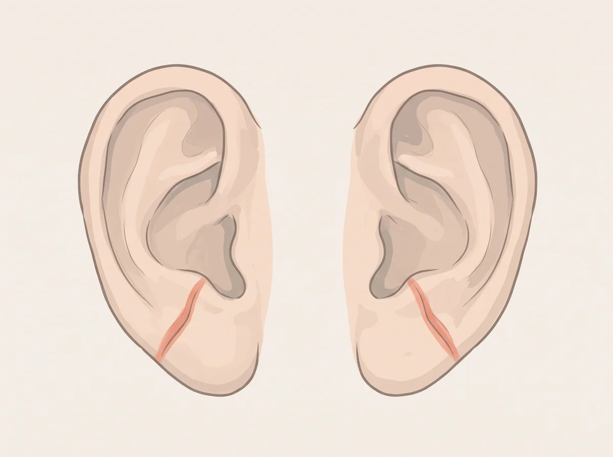
Illustration of diagonal earlobe crease. A clear fold runs at a slant across the fleshy earlobe, from near the face down toward the back edge of the lobe.  It can show on one ear or both.
Illustration, not a photograph
14

Diagonal earlobe crease

Frank’s sign (diagonal earlobe crease)

A diagonal earlobe crease is a faint, fun-to-spot heart clue

Mention at a checkup

Linked to
  • Coronary artery disease risk
  • Heart attack risk
What to look for
A clear fold runs at a slant across the fleshy earlobe, from near the face down toward the back edge of the lobe. It can show on one ear or both.
Why it happens
No one has proven the cause. Researchers think the crease shows wear on tiny blood vessels and stretchy tissue, the same kind of aging that stiffens heart arteries. Creases also get more common with age, which explains part of the link.
Other causes
Earlobe creases get more common with normal aging, and many people with a crease have healthy hearts.
How strong is the link
Real link A 35-year Danish study of 10,885 adults found that people with an earlobe crease had more heart disease and heart attacks, even after accounting for age and other known risk factors. A 2021 review of 13 studies found the crease is a poor test by itself: it misses many people with narrowed heart arteries and flags many people without them.
What to check
  • Blood pressure check
  • Cholesterol (lipid panel) and HbA1c blood tests
  • A 10-year heart risk estimate from your doctor
  1. Diagonal Earlobe Crease (Frank’s Sign) for Diagnosis of Coronary Artery Disease: A Systematic Review of Diagnostic Test Accuracy Studies · Journal of Clinical Medicine (PMC), 2021
  2. Visible Age-Related Signs and Risk of Ischemic Heart Disease in the General Population: A Prospective Cohort Study · Circulation, 2014
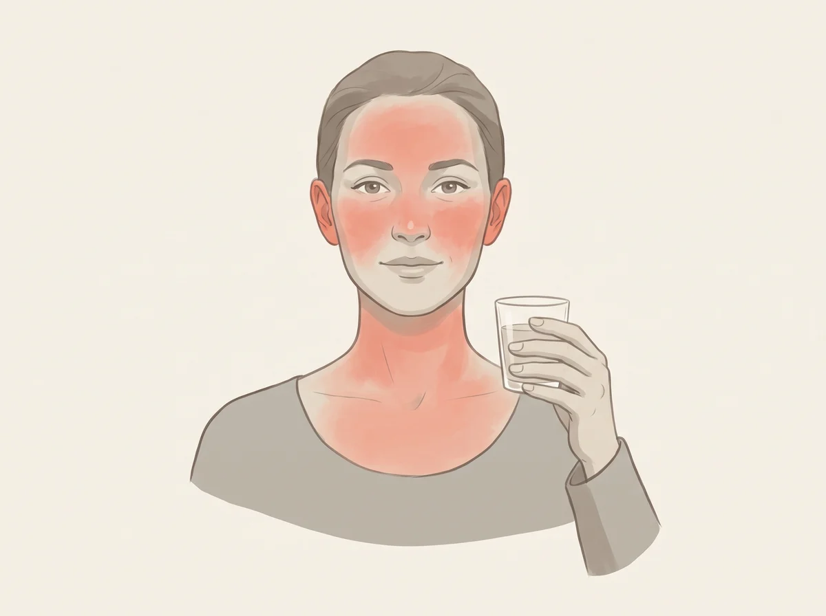
Illustration of face turns red after one drink. Within minutes of a small amount of alcohol, the face, neck, and sometimes chest turn red.  A racing heart or nausea often comes with it.
Illustration, not a photograph
15

Face turns red after one drink

Alcohol flushing response

A red face after one drink is a gene talking, and worth listening to

Mention at a checkup

Linked to
  • ALDH2 gene variant
  • Acetaldehyde buildup
  • Higher esophageal cancer risk with drinking
What to look for
Within minutes of a small amount of alcohol, the face, neck, and sometimes chest turn red. A racing heart or nausea often comes with it.
Why it happens
The body turns alcohol into acetaldehyde, a toxic chemical, and then an enzyme called ALDH2 clears it. Many people carry a gene variant (ALDH2*2) that makes this enzyme weak or inactive. Acetaldehyde then builds up and causes the flush.
Other causes
A red face after drinking can also come from a hot room or from rosacea, a skin condition that alcohol can set off. The gene variant is by far the most common cause in people of East Asian descent.
How strong is the link
Strong link About 36% of East Asians flush, and case-control studies found heavy drinkers with the variant had more than 10 times the risk of one type of esophageal cancer. Simply asking about flushing picked out the variant about 9 times out of 10 in Japanese adults.
What to check
  • Nothing to test. The flush already tells you what a gene test would.
  • Tell your doctor you flush, especially if you drink or smoke often.
  • If you have been a regular drinker for years, ask whether you need extra cancer screening.
  1. The Alcohol Flushing Response: An Unrecognized Risk Factor for Esophageal Cancer from Alcohol Consumption · PLOS Medicine, 2009
  2. The Alcohol Flush Response · Graphic Medicine Review, 2024

Mouth & teeth

16–19
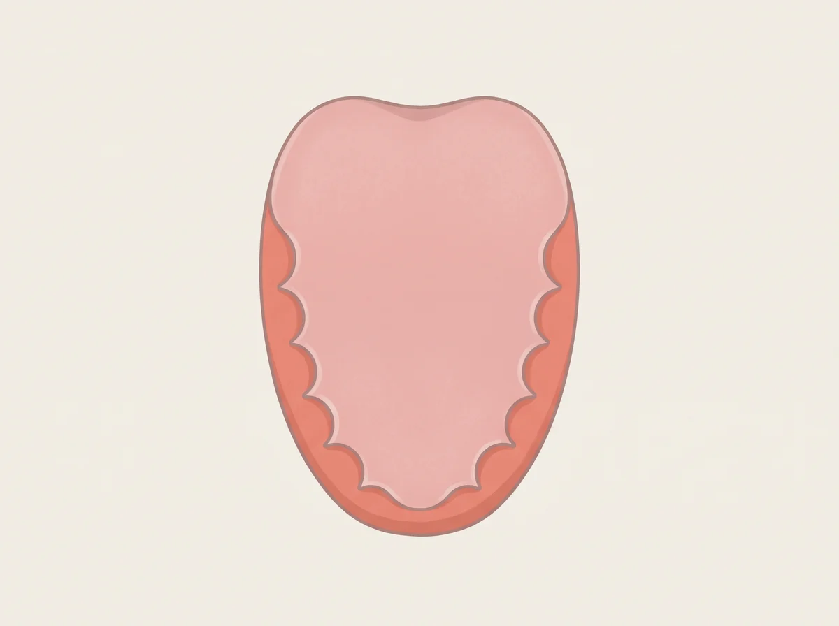
Illustration of wavy tooth marks along the tongue edges. The sides of the tongue have a wavy, rippled edge where the tongue has pressed against the teeth.  It is easiest to see when you stick your tongue out in a mirror.
Illustration, not a photograph
16

Wavy tooth marks along the tongue edges

Scalloped (crenated) tongue

Wavy tooth prints on your tongue edges can be a sleep clue

Mention at a checkup

Linked to
  • Obstructive sleep apnea
  • Underactive thyroid (hypothyroidism)
  • Enlarged tongue from other causes
  • Teeth clenching or grinding
What to look for
The sides of the tongue have a wavy, rippled edge where the tongue has pressed against the teeth. It is easiest to see when you stick your tongue out in a mirror.
Why it happens
The marks form when the tongue is wide for the space inside the teeth and rests against them for long stretches. A large tongue can also crowd the throat in sleep apnea, where the airway keeps closing during sleep. A low thyroid can make the tongue puff up because fluid-holding molecules collect in it.
Other causes
About 1 in 5 people without sleep apnea in one large study had tooth marks, often just from a naturally wide tongue. Rarer causes of a large tongue include amyloidosis and acromegaly.
How strong is the link
Early evidence In a 2025 study of 1,261 people, tooth marks showed up in 40% of those with sleep apnea and 21% of those without, so the sign misses most cases by itself. The popular link to clenching is weak: a small lab study found tongue width mattered more than how hard people pressed.
What to check
  • Sleep study (home test or overnight polysomnography)
  • TSH and free T4 blood tests
  • Dental exam for signs of grinding or clenching

Get seen soon Ask about a sleep study soon if you also snore loudly, stop breathing or gasp in your sleep, or feel sleepy while driving.

  1. Tongue scalloping as a sign of obstructive sleep apnea syndrome: Clinical and polysomnographic insights · Lung India (PMC), 2025
  2. Scalloped tongue: an additional, accessible and useful tool to detect severe obstructive sleep apnea? · Frontiers in Sleep, 2025
  3. Influence of tongue pressure and width on tongue indentation formation · Journal of Oral Rehabilitation (PubMed), 2007
  4. Macroglossia · StatPearls, NCBI Bookshelf, 2023
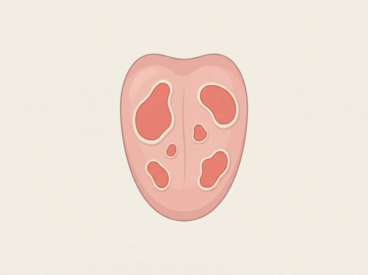
Illustration of map-like smooth patches on the tongue that move around. Smooth red patches with white or yellow wavy borders, like countries on a map.  The patches change shape and move to new spots over days or weeks.
Illustration, not a photograph
17

Map-like smooth patches on the tongue that move around

Geographic tongue (benign migratory glossitis)

A tongue that looks like a map, and the map keeps redrawing itself

Just a quirk

Linked to
  • A harmless quirk
  • Psoriasis
  • Allergies and eczema
  • Family history
What to look for
Smooth red patches with white or yellow wavy borders, like countries on a map. The patches change shape and move to new spots over days or weeks.
Why it happens
In the patches, the tongue loses its tiny bumps for a while, so the red surface shows. Doctors do not know why this happens. It tends to run in families and flares with stress and hormone changes.
Other causes
It shows up more often in people with psoriasis, allergies, asthma, or eczema. It affects about 1 to 2.5% of people.
How strong is the link
Real link A 2019 review of 11 studies found map tongue in about 10% of people with psoriasis, compared with about 5% of people without it. The tongue pattern itself is harmless.
What to check
  • Nothing to test.
  • If it stings with spicy or sour food, mention it to your dentist.
  • If you also have scaly, itchy skin patches, ask your doctor about psoriasis.
  1. Geographic Tongue · StatPearls, NCBI Bookshelf, 2023
  2. Association between geographic tongue and psoriasis: A systematic review and meta-analyses · Journal of Oral Pathology & Medicine, 2019
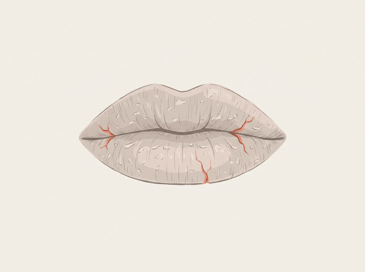
Illustration of lips that stay dry and cracked. Lips that feel tight, flake, peel, or crack and do not get better with a few days of lip balm.
Illustration, not a photograph
18

Lips that stay dry and cracked

Cheilitis

Licking chapped lips makes them worse, and other lip facts

Just a quirk

Linked to
  • Licking your lips
  • Dry, cold, or windy weather
  • Acne pills like isotretinoin
  • Low iron or B vitamins (less common)
What to look for
Lips that feel tight, flake, peel, or crack and do not get better with a few days of lip balm.
Why it happens
Licking feels like it helps, but saliva evaporates and leaves lips drier than before. Cold, hot, or dry air, wind, and sunburn dry lips out too. Isotretinoin, a strong acne pill, dries the lips in about 9 of 10 people who take it.
Other causes
An allergy to a lip product, other retinoid medicines, and low iron or B vitamins can cause lips that stay cracked. Thyroid problems and some autoimmune conditions can too.
How strong is the link
Real link The effect of isotretinoin is well measured, and dermatologists agree on lip licking and weather. Vitamin and iron shortfalls are a real but less common cause.
What to check
  • Nothing to test at first. Try 2 to 3 weeks of a plain, unflavored lip balm and no licking.
  • Still cracked after that: ask a doctor about allergies to lip products, your medicines, and iron and B vitamin levels.
  • A rough, scaly spot on the lower lip that will not heal needs a dermatologist’s look.
  1. 7 dermatologists’ tips for healing dry, chapped lips · American Academy of Dermatology
  2. Chapped Lips (Cheilitis) · Cleveland Clinic, 2024
  3. Isotretinoin · StatPearls, NCBI Bookshelf, 2025
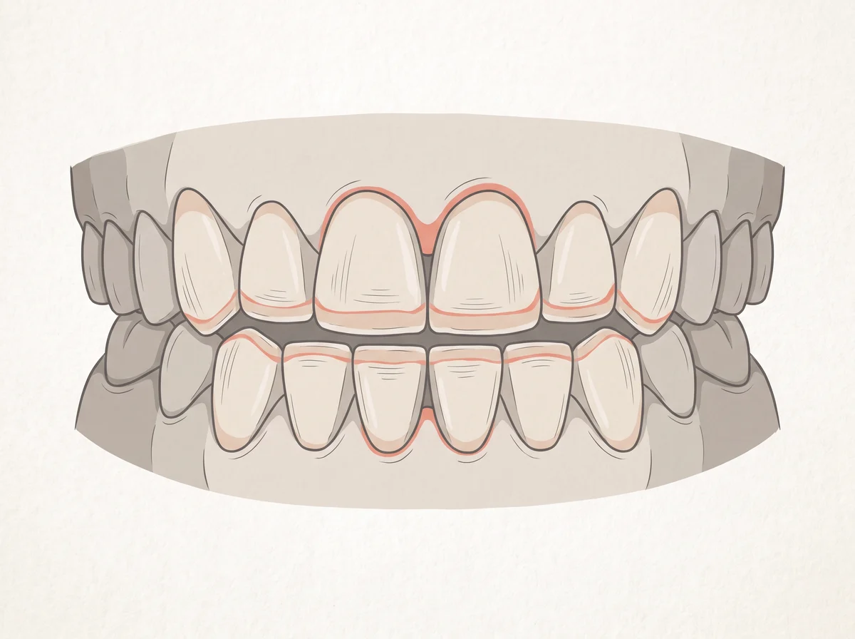
Illustration of flat, worn-down teeth from grinding. The biting edges of your front teeth look flat, even, or chipped instead of slightly rounded.  Teeth may feel sensitive, and your jaw may ache in the morning.
Illustration, not a photograph
19

Flat, worn-down teeth from grinding

Tooth wear from bruxism

Flat front teeth can reveal a grinding habit you never noticed

Mention at a checkup

Linked to
  • Stress and anxiety
  • Teeth grinding in your sleep
  • Sleep apnea
  • Caffeine, alcohol, and smoking
What to look for
The biting edges of your front teeth look flat, even, or chipped instead of slightly rounded. Teeth may feel sensitive, and your jaw may ache in the morning.
Why it happens
Bruxism means clenching or grinding your teeth, awake or asleep. Night grinding wears enamel off the biting edges, and most people do not know they do it. Stress is the most common trigger, and about half of adults with obstructive sleep apnea also grind in their sleep.
Other causes
A bite that does not line up, or missing or crooked teeth, can lead to grinding. Some medicines, such as SSRI antidepressants, can too. People who smoke or drink alcohol or coffee are about twice as likely to grind.
How strong is the link
Real link Sleep grinding affects about 8 to 10% of adults, and the links to stress and sleep apnea are well described. Grinding is diagnosed mainly from tooth wear and what a bed partner notices, so exact numbers vary.
What to check
  • Ask your dentist to check for wear at your next cleaning.
  • Ask whether a night guard makes sense for you.
  • If you snore loudly or wake up tired, ask your doctor about a sleep apnea test.
  1. Bruxism Management · StatPearls, NCBI Bookshelf, 2024
  2. Teeth Grinding · MouthHealthy, American Dental Association

Skin

20–26
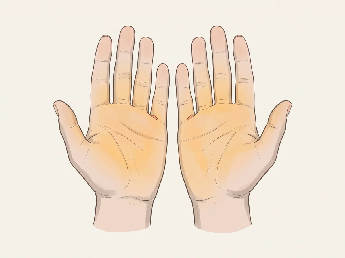
Illustration of orange-tinted palms and soles. A yellow-orange tint that is strongest on the palms, soles, and sometimes the tip of the nose.  The whites of the eyes stay white.
Illustration, not a photograph
20

Orange-tinted palms and soles

Carotenemia (carotenoderma)

Orange palms usually mean you have been loving your carrots

Just a quirk

Linked to
  • High-carotene diet
  • Hypothyroidism
  • Diabetes
  • Liver or kidney disease
What to look for
A yellow-orange tint that is strongest on the palms, soles, and sometimes the tip of the nose. The whites of the eyes stay white.
Why it happens
Beta-carotene is the orange pigment in carrots, sweet potatoes, and squash. When blood levels run high, it collects in the thick outer skin of the palms and soles. An underactive thyroid, diabetes, and liver or kidney disease can slow how the body turns carotene into vitamin A, so it builds up even on a normal diet.
Other causes
Eating a lot of carrots, squash, sweet potatoes, papaya, mango, or leafy greens is the most common cause. Babies on puréed vegetables often get it, and it is harmless.
How strong is the link
Real link Carotenemia is well described and harmless in itself, and diet is the usual cause. Hypothyroidism, diabetes, and liver or kidney disease are known but less common causes, which is why doctors screen for them when diet does not explain it.
What to check
  • Look at the whites of the eyes (yellow eyes mean jaundice)
  • TSH blood test
  • A1c or fasting blood sugar
  • Liver panel with bilirubin
  • Kidney function (creatinine)
  • Serum carotene level

Get seen soon If the whites of your eyes turn yellow too, see a doctor soon. Yellow eyes point to the liver or blood, which is a different sign.

  1. Carotenemia · StatPearls, NCBI Bookshelf, 2023
  2. Carotenemia: A Case Report · Cureus (PMC), 2019
  3. Decoding Hypercarotenemia: Integrating Pathophysiology, Clinical Recognition, and Precision Management · Cureus (PMC), 2025
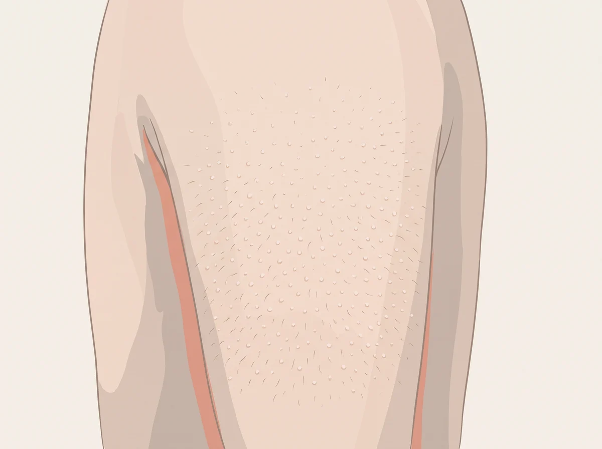
Illustration of tiny rough bumps on the backs of the arms. Small, rough bumps that feel like sandpaper, mostly on the backs of the upper arms.  They can also show up on the thighs, butt, or cheeks, and may look skin-colored, pink, or red.
Illustration, not a photograph
21

Tiny rough bumps on the backs of the arms

Keratosis pilaris (chicken skin)

Chicken skin on your arms is common and harmless

Just a quirk

Linked to
  • Family genes
  • Dry skin
  • Eczema (atopic dermatitis)
  • Dry winter air
What to look for
Small, rough bumps that feel like sandpaper, mostly on the backs of the upper arms. They can also show up on the thighs, butt, or cheeks, and may look skin-colored, pink, or red.
Why it happens
Your skin makes a tough protein called keratin. In keratosis pilaris, extra keratin plugs the opening of each hair follicle and forms a tiny hard bump. It runs in families and is tied to changes in the gene for filaggrin, a protein that helps skin hold in water.
Other causes
It is more common in people with eczema, ichthyosis vulgaris (very dry, scaly skin), or obesity. About half of people see it get better in summer and worse in winter, when the air is dry.
How strong is the link
Strong link About 50 to 80 percent of teens and about 40 percent of adults have it, so it acts more like a skin type than a disease. Major reviews do not list low vitamin A as a cause. A look-alike rash called phrynoderma shows up with severe malnutrition, which is a different problem.
What to check
  • Nothing to test. Mention it if the bumps spread fast, itch a lot, or show up along with weight loss or a very poor diet.
  1. Keratosis Pilaris · StatPearls, NCBI Bookshelf, 2023
  2. Keratosis pilaris and filaggrin loss-of-function mutations in patients with atopic dermatitis: Results of a Finnish cross-sectional study · Journal of Dermatology, 2022
  3. Phrynoderma: a manifestation of vitamin A deficiency?... The rest of the story · Pediatric Dermatology, 2005
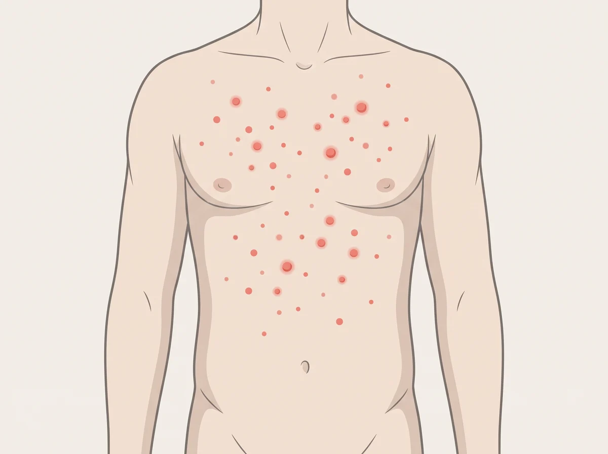
Illustration of tiny bright red dots on the chest or belly. Round spots, light red to dark red, from pinhead size up to about 4 millimeters across.  They are smooth or slightly raised and show up most on the chest, belly, back, arms, and legs.
Illustration, not a photograph
22

Tiny bright red dots on the chest or belly

Cherry angioma (cherry hemangioma)

Little red dots are a normal part of getting older

Just a quirk

Linked to
  • Normal aging
  • Family genes
  • Pregnancy hormones
  • Rarely, certain chemicals
What to look for
Round spots, light red to dark red, from pinhead size up to about 4 millimeters across. They are smooth or slightly raised and show up most on the chest, belly, back, arms, and legs.
Why it happens
Each dot is a small cluster of tiny blood vessels that grew near the surface of the skin. Scientists have found gene changes, in genes called GNAQ and GNA11, inside the spots. You tend to get more of them with each decade of life.
Other causes
Hormone shifts during pregnancy can bring on new spots. Rarely, exposure to certain chemicals, such as nitrogen mustard or bromides, is linked to them.
How strong is the link
Strong link About half of adults over 30 have at least one, and about 75 percent of people over 75 do. They have no cancer risk.
What to check
  • Nothing to test. Show a doctor any spot that bleeds often, changes shape or color, or looks different from your other spots.

Get seen soon Get checked soon if many new red spots appear all at once and you also feel sick or have swollen glands.

  1. Cherry Hemangioma · StatPearls, NCBI Bookshelf, 2023
  2. Cherry Angioma · Cleveland Clinic
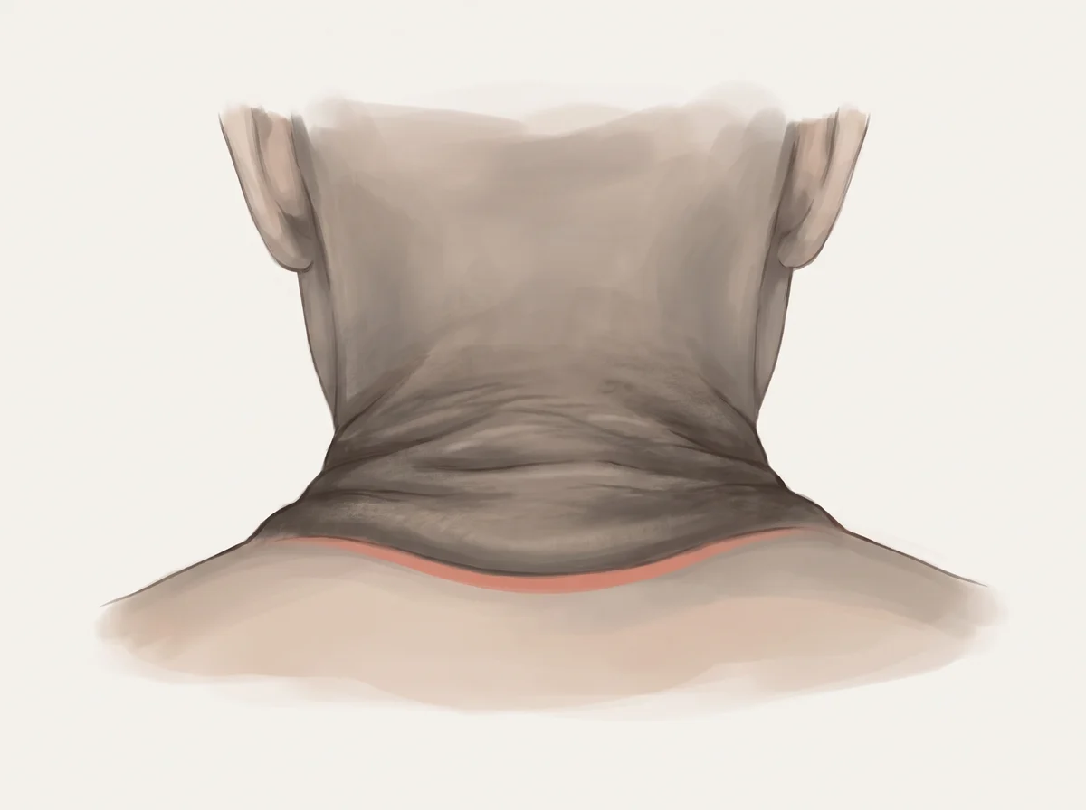
Illustration of dark, velvety skin on the neck or armpits. Skin in body folds, most often the back and sides of the neck or the armpits, turns darker and thicker with a soft, velvety feel.  Elbows and knuckles can change too.
Illustration, not a photograph
23

Dark, velvety skin on the neck or armpits

Acanthosis nigricans

Soft, dark skin in body folds is a nudge to check blood sugar

Mention at a checkup

Linked to
  • Insulin resistance
  • Prediabetes or type 2 diabetes
  • Polycystic ovary syndrome (PCOS)
  • Rarely, internal cancer
What to look for
Skin in body folds, most often the back and sides of the neck or the armpits, turns darker and thicker with a soft, velvety feel. Elbows and knuckles can change too. It can look like a shadow that does not wash off.
Why it happens
When the body resists insulin, the pancreas makes extra insulin to keep blood sugar normal. High insulin acts like a growth signal for skin cells, so the skin in folds thickens and darkens.
Other causes
It runs in some families and is more common in some ethnic groups. Some medicines, including steroids, birth control pills, and niacin, can also cause it.
How strong is the link
Strong link Obesity and insulin resistance cause most cases, and the American Academy of Dermatology notes it can be the first visible sign of prediabetes. The skin often fades when weight and insulin levels improve.
What to check
  • HbA1c
  • Fasting glucose, plus fasting insulin if your doctor uses it
  • Lipid panel
  • Hormone tests for PCOS, such as testosterone, if periods are irregular

Get seen soon See a doctor soon if dark patches appear suddenly, spread fast, show up on the palms or inside the mouth, or come with weight loss you cannot explain. That rare pattern can signal cancer and needs a closer look.

  1. Acanthosis Nigricans · StatPearls, NCBI Bookshelf, 2023
  2. Acanthosis nigricans: Who gets and causes · American Academy of Dermatology
  3. Diabetes: 10 warning signs that can appear on your skin · American Academy of Dermatology
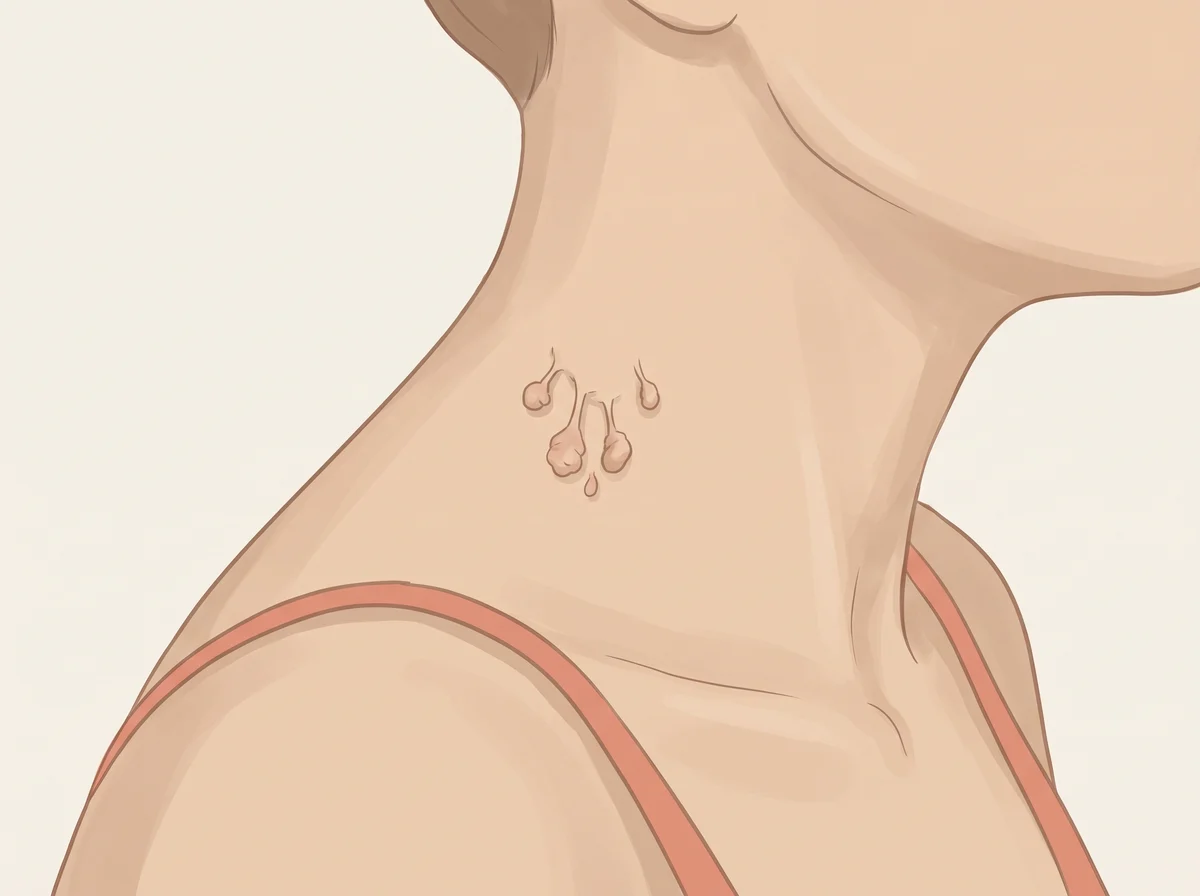
Illustration of many skin tags on the neck, armpits, or eyelids. Skin tags are small, soft, skin-colored or brown bits of skin that often hang from a thin stalk.  They gather where skin rubs, such as the neck, armpits, groin, and eyelids.
Illustration, not a photograph
24

Many skin tags on the neck, armpits, or eyelids

Acrochordons

One skin tag is nothing, but a crowd of them hints at blood sugar

Mention at a checkup

Linked to
  • Insulin resistance
  • Type 2 diabetes
  • Obesity
  • Metabolic syndrome
What to look for
Skin tags are small, soft, skin-colored or brown bits of skin that often hang from a thin stalk. They gather where skin rubs, such as the neck, armpits, groin, and eyelids.
Why it happens
Rubbing in skin folds seems to start them. High insulin levels act as a growth signal for skin, which may explain why people with insulin resistance grow more of them.
Other causes
About half of adults get at least one skin tag, and they become more common with age, pregnancy, and weight gain. One or two tags by themselves mean very little.
How strong is the link
Real link In a study of 110 people with several skin tags, 52% had diabetes, compared with 10% of matched people without tags. Skin tags are so common that the link matters most when you have many of them or they appear quickly.
What to check
  • HbA1c
  • Fasting glucose
  • Lipid panel
  • Blood pressure and waist measurement

Get seen soon See a doctor if many new tags appear in a short time, or if any growth bleeds, changes color, or grows quickly.

  1. Skin Tag (Acrochordon) · StatPearls, NCBI Bookshelf, 2025
  2. Acrochordons as a Cutaneous Sign of Metabolic Syndrome: A Case-Control Study · Annals of Medical and Health Sciences Research (PMC), 2014
  3. Skin tags: Why they develop, and how to remove them · American Academy of Dermatology
  4. Diabetes: 10 warning signs that can appear on your skin · American Academy of Dermatology
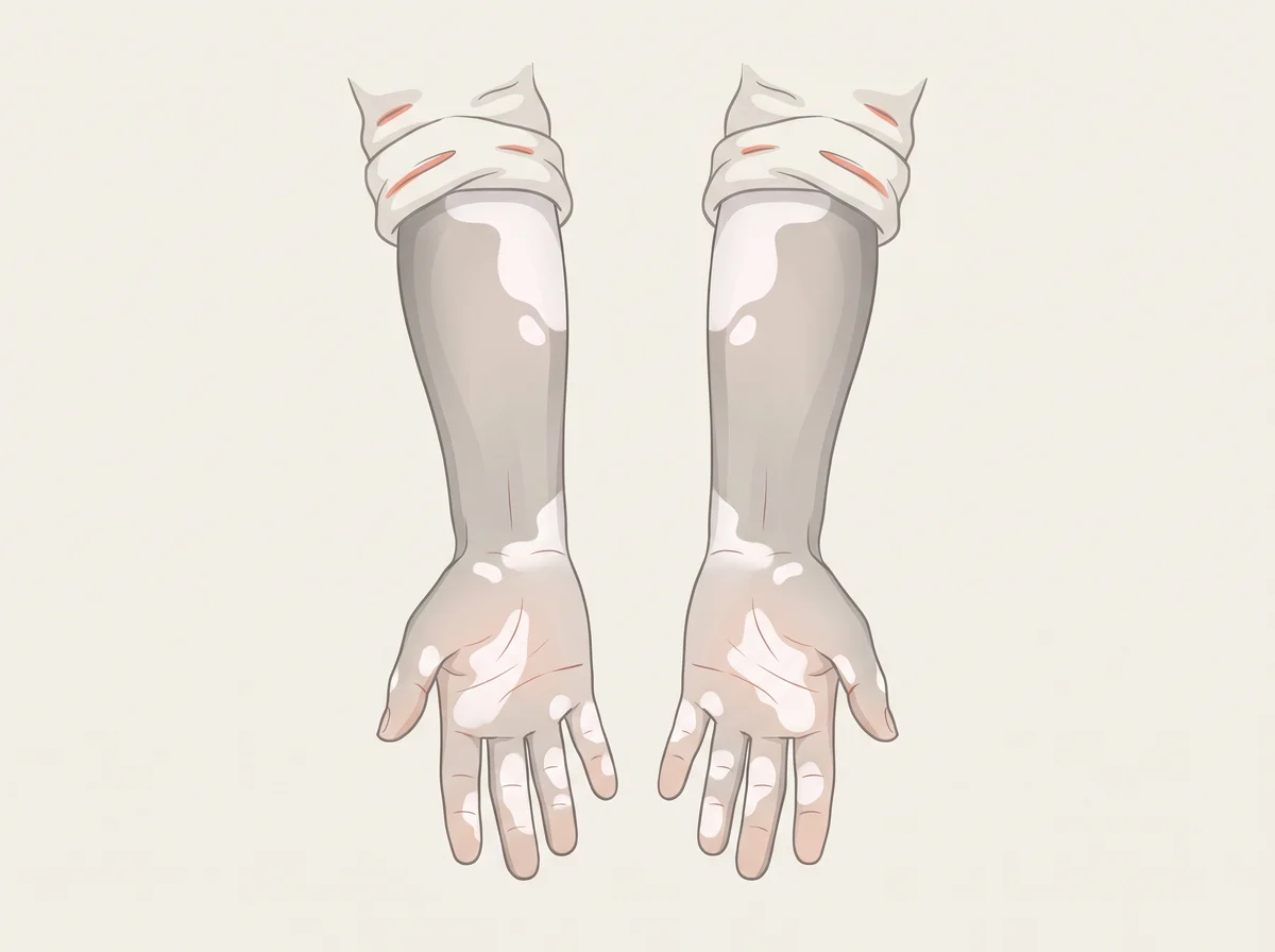
Illustration of white patches of skin that lost their color. Milky white, sharply edged patches with no pigment, often on the hands, fingers, face, around the mouth and eyes, or on the elbows and knees.  The patches often show up on both sides of the body.
Illustration, not a photograph
25

White patches of skin that lost their color

Vitiligo

Vitiligo patches come with a good excuse to check your thyroid

Mention at a checkup

Linked to
  • Autoimmune thyroid disease
  • Type 1 diabetes
  • Pernicious anemia (low B12)
What to look for
Milky white, sharply edged patches with no pigment, often on the hands, fingers, face, around the mouth and eyes, or on the elbows and knees. The patches often show up on both sides of the body.
Why it happens
Vitiligo is an autoimmune condition, which means the immune system mistakes some of the body’s own cells for invaders. Here it targets melanocytes, the cells that make skin pigment. An immune system that does this in one place is more likely to do it elsewhere, such as the thyroid, the pancreas, or the stomach cells that help absorb vitamin B12.
Other causes
Pale patches can also come from a yeast infection called tinea versicolor, from eczema that has healed, from old burns or scars, or from pityriasis alba, a harmless dry patch common in children.
How strong is the link
Real link Large reviews find autoimmune thyroid disease in about 14% of people with vitiligo, about 2.5 to 6 times the risk in people without it. Type 1 diabetes (about 1.8%) and pernicious anemia (about 1.7%) are less common. Most people with vitiligo do not have another autoimmune disease.
What to check
  • TSH blood test
  • TPO (thyroid peroxidase) antibodies
  • A1c or fasting blood sugar
  • Complete blood count and vitamin B12 level
  • Wood’s lamp skin exam by a dermatologist

Get seen soon Book a visit within a few weeks if the patches come with tiredness, weight change, heavy thirst, or numb or tingling hands and feet.

  1. Vitiligo and thyroid disease: a systematic review and meta-analysis · European Journal of Dermatology (PubMed), 2018
  2. The prevalence of thyroid disease in patients with vitiligo: a systematic review · British Journal of Dermatology (PubMed), 2012
  3. Prevalence and Association of Autoimmune Comorbidities Among Adults with Vitiligo: A Systematic Literature Review and Meta-analysis of USA-Based Studies · Dermatology and Therapy (PMC), 2025
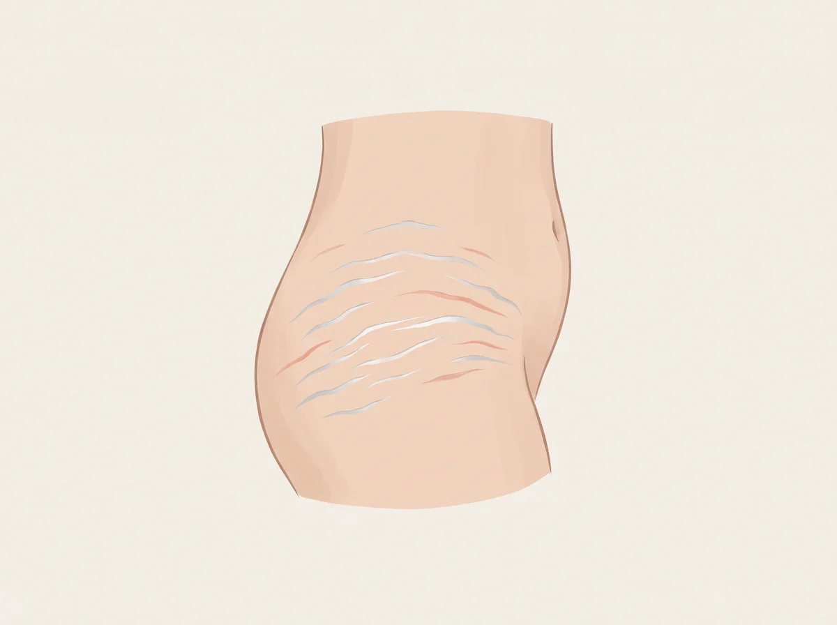
Illustration of thin silvery stretch marks. Thin lines that look paler or shinier than the skin around them, often on the hips, thighs, belly, breasts, butt, or lower back.  New marks start pink, red, or purple and fade to silver or white over months to years.
Illustration, not a photograph
26

Thin silvery stretch marks

Striae distensae (called striae albae once they fade)

Silver stripes are skin that grew faster than it could stretch

Just a quirk

Linked to
  • Growth spurts
  • Pregnancy
  • Fast weight or muscle gain
  • Steroid medicines
What to look for
Thin lines that look paler or shinier than the skin around them, often on the hips, thighs, belly, breasts, butt, or lower back. New marks start pink, red, or purple and fade to silver or white over months to years.
Why it happens
The middle layer of skin, called the dermis, holds springy fibers called collagen and elastin. When skin stretches fast, or hormones shift, those fibers break and heal like a thin scar. The fresh scar looks pink or red, then slowly fades.
Other causes
Long use of steroid pills or creams can cause them. Family history, being female, and being overweight raise the odds. Rarely, they come from Cushing syndrome, where the body makes too much of the stress hormone cortisol.
How strong is the link
Strong link Studies find stretch marks in 6 to 86 percent of teens and 43 to 88 percent of pregnant people. Thin pale marks from puberty, pregnancy, or weight change are a normal scar and not a health problem.
What to check
  • Nothing to test for thin silvery marks.
  • Ask your doctor about new marks that are wide and purple when you have no clear reason, such as pregnancy, a growth spurt, or weight gain.

Get seen soon Book a visit if wide purple stretch marks appear along with a rounder face, weight gain around the belly and upper back, easy bruising, or weak muscles.

  1. Striae Distensae · StatPearls, NCBI Bookshelf, 2025
  2. Stretch marks · NHS
  3. Cushing’s Syndrome · National Institute of Diabetes and Digestive and Kidney Diseases (NIDDK)

Hands & nails

27–42
Illustration of fingers that turn white, then blue, in the cold. In the cold or under stress, one or more fingers turn a sharp white, then blue, then red as they warm up.  The fingers feel numb or cold during the spell and may tingle or throb afterward.
Illustration, not a photograph
27

Fingers that turn white, then blue, in the cold

Raynaud phenomenon

Fingers that go ghost-white in the cold are usually just touchy

Mention at a checkup

Linked to
  • Raynaud phenomenon
  • Scleroderma (systemic sclerosis)
  • Lupus
  • Thyroid disease
What to look for
In the cold or under stress, one or more fingers turn a sharp white, then blue, then red as they warm up. The fingers feel numb or cold during the spell and may tingle or throb afterward.
Why it happens
Small arteries in the fingers clamp down too hard in response to cold or stress, and blood flow drops for a while. The finger goes white from too little blood, blue as the trapped blood uses up its oxygen, and red when blood rushes back. In the less common secondary form, a disease such as scleroderma damages these small vessels and makes the spasms worse.
Other causes
Most people have primary Raynaud, which has no underlying disease and is more common in young women and in families. Beta-blocker drugs, some migraine and ADHD medicines, and years of using vibrating tools can also cause it.
How strong is the link
Strong link The color change is a well-defined sign of Raynaud phenomenon, and most cases are the harmless primary type. The link to autoimmune disease is strong in a smaller group. Over 95 percent of people with scleroderma have Raynaud, and in one common form it can start years before the disease reaches the organs, which is why a new case is worth one checkup.
What to check
  • Nailfold capillaroscopy (a magnified look at the tiny vessels at the base of the nails)
  • Antinuclear antibody (ANA) blood test
  • ESR or CRP for inflammation
  • TSH (thyroid stimulating hormone)
  • Complete blood count (CBC)

Get seen soon See a doctor soon if Raynaud starts after age 30, affects only one hand, involves the thumb, or causes sores on the fingertips.

  1. Raynaud’s Phenomenon · National Institute of Arthritis and Musculoskeletal and Skin Diseases (NIAMS), 2024
  2. Raynaud Disease · StatPearls, NCBI Bookshelf, 2023
  3. Systemic Sclerosis (Scleroderma) · StatPearls, NCBI Bookshelf, 2024
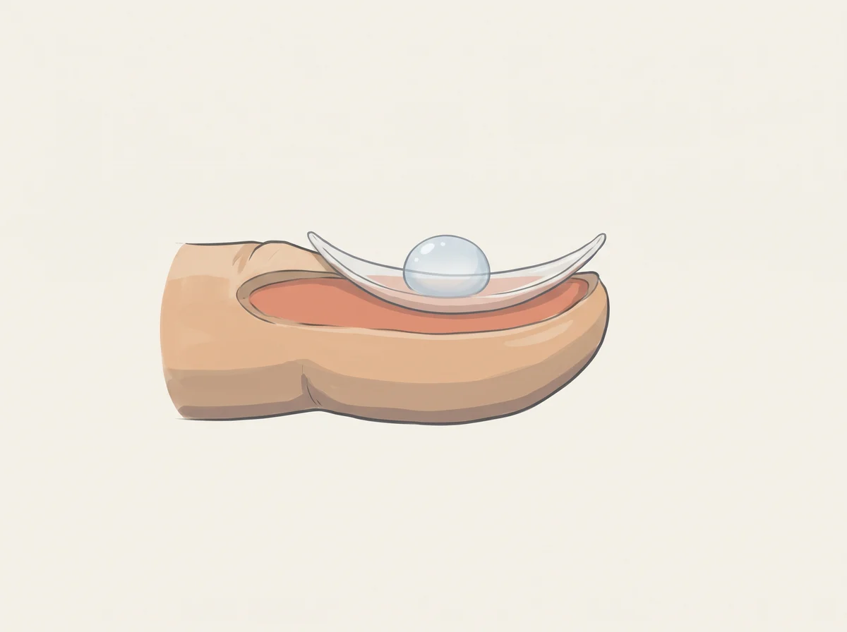
Illustration of palms that sweat for no reason. Both palms get wet, sometimes dripping, even when you feel cool and calm.  The soles or armpits may sweat too.
Illustration, not a photograph
28

Palms that sweat for no reason

Primary focal hyperhidrosis

Damp handshakes often run in the family

Just a quirk

Linked to
  • Family genes (primary hyperhidrosis)
  • Stress or nerves
  • Overactive thyroid
  • Some medicines
What to look for
Both palms get wet, sometimes dripping, even when you feel cool and calm. The soles or armpits may sweat too. It usually starts before age 25 and eases or stops while you sleep.
Why it happens
Nerves tell sweat glands when to switch on. In primary hyperhidrosis, those nerve signals run too strong, so the glands in the hands make far more sweat than your body needs to cool down. Stress, heat, and caffeine can turn it up.
Other causes
Sweating all over the body, heavy night sweats, or sweating that starts later in life points to other causes. These include an overactive thyroid, diabetes, menopause, and some medicines, such as the antidepressant sertraline.
How strong is the link
Strong link About 3 percent of people in the US have hyperhidrosis, and it clusters in families. Sweaty palms on both hands that began before age 25 and ease during sleep fit the harmless primary type.
What to check
  • Nothing to test if it started young and only affects your hands, feet, or armpits.
  • Ask for a thyroid blood test if the sweating started as an adult or comes with weight loss, a racing heart, or shaky hands.
  • Ask a doctor about treatments if it gets in the way of daily life.

Get seen soon See a doctor soon if you start having heavy sweats at night or all over your body, especially with a fever or weight loss you cannot explain.

  1. Hyperhidrosis · StatPearls, NCBI Bookshelf, 2022
  2. Hyperhidrosis · Cleveland Clinic, 2023
  3. Hyperthyroidism (Overactive Thyroid) · National Institute of Diabetes and Digestive and Kidney Diseases (NIDDK), 2021
Illustration of ring finger longer than index finger. Hold your hand flat, palm up.  Measure your index finger (2D) and ring finger (4D) from the crease at the base to the tip.
Illustration, not a photograph
29

Ring finger longer than index finger

2D:4D digit ratio

Finger length ratios make great trivia and weak health clues

Just a quirk

Linked to
  • Sex (men tend to have a lower ratio)
  • Possibly hormones before birth
  • Weak links to traits and health
What to look for
Hold your hand flat, palm up. Measure your index finger (2D) and ring finger (4D) from the crease at the base to the tip. If the ring finger is longer, your ratio is below 1, a pattern more common in men.
Why it happens
Finger length is set before birth. Scientists proposed that more testosterone in the womb makes the ring finger a bit longer than the index finger. Men do have a lower ratio on average, and the gap between men and women is a bit bigger on the right hand.
Other causes
How you measure changes the number, whether you use a ruler, a photo, or a scan. Your right and left hands can also give different ratios.
How strong is the link
Early evidence People with congenital adrenal hyperplasia, who make extra male hormones before birth, have slightly lower ratios on average. But a 2024 review of 54 studies found no clear link between the ratio and testosterone measured before birth or in adults, so it cannot tell you much about your own hormones, personality, or health.
What to check
  • Nothing to test. Treat it as a fun fact about your hands.
  1. Relationship between the 2D:4D and prenatal testosterone, adult level testosterone, and testosterone change: Meta-analysis of 54 studies · American Journal of Biological Anthropology, 2024
  2. Meta-analysis of digit ratio 2D:4D shows greater sex difference in the right hand · American Journal of Human Biology, 2010
  3. Digit ratio (2D:4D) and congenital adrenal hyperplasia (CAH): Systematic literature review and meta-analysis · Hormones and Behavior, 2020
Illustration of thumbs or fingers that bend back further than most. Your thumb can bend back to touch your forearm, your pinky bends back past 90 degrees, or your elbows and knees bend past straight.  Doctors count these moves on a 9-point check called the Beighton score.
Illustration, not a photograph
30

Thumbs or fingers that bend back further than most

Joint hypermobility (often called double-jointed)

Bendy thumbs come from stretchy ligaments you likely inherited

Just a quirk

Linked to
  • Family genes
  • Hypermobility spectrum disorder
  • POTS (dizziness when standing)
  • Anxiety
What to look for
Your thumb can bend back to touch your forearm, your pinky bends back past 90 degrees, or your elbows and knees bend past straight. Doctors count these moves on a 9-point check called the Beighton score.
Why it happens
Ligaments are the tough bands that hold joints together. Some people inherit ligaments made with a looser type of collagen, so their joints move further than usual. It shows up most in children and young people and often lessens with age.
Other causes
Rarely, very loose joints are part of an inherited condition of connective tissue, the body’s support material. Examples include Ehlers-Danlos syndrome and Marfan syndrome.
How strong is the link
Real link Doctors estimate about 3 percent of people have joint hypermobility syndrome, where loose joints come with symptoms. A 2014 review found these people had about 4 times the odds of anxiety, and people with hypermobility report POTS-type dizziness more often than others.
What to check
  • Nothing to test if your joints feel fine.
  • Mention it at a checkup if you also get frequent sprains or dislocations, lasting joint pain, constant tiredness, or dizziness when you stand up.
  • Ask about the Beighton score if a doctor is checking you for a hypermobility disorder.
  1. Beighton Score: How It’s Used to Measure Joint Hypermobility · Cleveland Clinic, 2022
  2. Joint hypermobility syndrome · NHS, 2023
  3. The relationship between benign joint hypermobility syndrome and psychological distress: a systematic review and meta-analysis · Rheumatology (Oxford), 2014
  4. Postural tachycardia syndrome and other forms of orthostatic intolerance in Ehlers-Danlos syndrome · Autonomic Neuroscience: Basic and Clinical, 2018
Illustration of bony knobs on the end finger joints. Hard, pea-sized bumps on the finger joint closest to the nail, often one on each side of the joint.  Similar bumps on the middle finger joints are called Bouchard’s nodes.
Illustration, not a photograph
31

Bony knobs on the end finger joints

Heberden’s nodes

Pea-sized bumps by your fingernails mark common hand arthritis

Just a quirk

Linked to
  • Hand osteoarthritis
  • Family genes
  • Getting older
  • Being female
What to look for
Hard, pea-sized bumps on the finger joint closest to the nail, often one on each side of the joint. Similar bumps on the middle finger joints are called Bouchard’s nodes.
Why it happens
They are a sign of osteoarthritis, the wear-and-tear type of arthritis. As the smooth cartilage inside the joint breaks down, the body builds new bone at the joint edges, and that bone forms the knobs. They can ache, swell, or feel stiff while they form.
Other causes
They run in families. A study of 931 family members found a pattern of inheritance that differs between women and men, and they are far more common in women.
How strong is the link
Strong link Heberden’s nodes are a classic sign doctors use to spot hand osteoarthritis. About half of women and 1 in 4 men have osteoarthritis in the hands by age 85.
What to check
  • Nothing to test if they do not bother you.
  • Mention them at a checkup if they hurt, swell, or make buttons and jar lids hard to manage.
  • Ask about hand therapy exercises, splints, or heat for sore joints.
  1. Heberden’s Nodes · Cleveland Clinic, 2021
  2. Osteoarthritis · StatPearls, NCBI Bookshelf, 2023
  3. Investigations in generalized osteoarthritis. Part 1: genetic study of Heberden’s nodes · Osteoarthritis and Cartilage, 2006
Illustration of hair on the middle part of your fingers. Look at the backs of your fingers, between the two knuckles.  Some people have a few hairs on that middle segment of one or more fingers, and others have none.
Illustration, not a photograph
32

Hair on the middle part of your fingers

Mid-digital (midphalangeal) hair

Hair on your middle finger segments is harmless trivia

Just a quirk

Linked to
  • Family genes
  • Ancestry
  • Wear from hand work
What to look for
Look at the backs of your fingers, between the two knuckles. Some people have a few hairs on that middle segment of one or more fingers, and others have none.
Why it happens
Whether hair grows there depends partly on your genes. Genetics classes once taught it as a simple one-gene trait, but family and twin studies do not fit that neat pattern. Hand work that rubs the hairs away may also play a part.
Other causes
How common it is changes a lot with ancestry. Studies report it in about 2 percent of Inuit people and up to about 75 percent of people with northern European roots.
How strong is the link
Real link A 1942 study found that a child rarely has mid-finger hair on more fingers than the hairier parent, but later family and twin studies gave mixed results. Research on it covers inheritance and ancestry, and it has no known health meaning.
What to check
  • Nothing to test. Count the hairy fingers in your family for fun.
  1. Myths of Human Genetics: Mid-digital Hair · John H. McDonald, University of Delaware, 2011
  2. The Incidence and Mendelian Transmission of Mid-Digital Hair in Man · Journal of Heredity, 1942
Illustration of dark patches over the knuckles. Skin over the knuckles turns clearly darker than the rest of the hand, often on both hands.  With high insulin the skin can also feel thick and velvety, and similar dark patches may show up on the neck or armpits.
Illustration, not a photograph
33

Dark patches over the knuckles

Knuckle hyperpigmentation; acanthosis nigricans of the knuckles

Darker knuckles can be a small clue about B12 or insulin

Mention at a checkup

Linked to
  • Insulin resistance
  • Vitamin B12 deficiency
  • Prediabetes
What to look for
Skin over the knuckles turns clearly darker than the rest of the hand, often on both hands. With high insulin the skin can also feel thick and velvety, and similar dark patches may show up on the neck or armpits.
Why it happens
High insulin levels push skin cells to multiply, which thickens and darkens the skin in spots like the knuckles and neck. With low B12, the skin makes extra pigment for reasons doctors do not fully understand, and it fades once B12 levels recover.
Other causes
Darker skin over the knuckles is common and normal in many people with brown or black skin. Friction, sun, and Addison disease (low adrenal hormones) can also darken knuckles.
How strong is the link
Early evidence The B12 link comes mainly from case reports, where the dark patches faded after treatment, so this is weak evidence. For insulin resistance, one study of 294 young adults found higher insulin in people with dark, velvety knuckles, but the sign missed about 3 in 10 cases and flagged many people without it.
What to check
  • Serum vitamin B12, with methylmalonic acid (MMA) or homocysteine if B12 is borderline
  • Complete blood count (CBC) with MCV (red cell size)
  • HbA1c and fasting glucose
  • Fasting insulin (to estimate insulin resistance)
  • Ask about metformin, acid-reducing drugs, or a vegan diet, which raise B12 risk

Get seen soon See a doctor soon if dark knuckles come with numbness or tingling in the hands or feet, balance trouble, or dark velvety patches that appear over a few weeks.

  1. Knuckle Pigmentation as an Early Cutaneous Sign of Vitamin B12 Deficiency: A Case Report · Journal of the Nepal Medical Association (Srivastava, Choudhary), 2020
  2. Acanthosis nigricans in the knuckles: An early, accessible, straightforward, and sensitive clinical tool to predict insulin resistance · Dermato-Endocrinology (González-Saldivar et al.), 2018
  3. Acanthosis Nigricans · StatPearls, NCBI Bookshelf, 2023
  4. Vitamin B12 Deficiency · StatPearls, NCBI Bookshelf, 2024
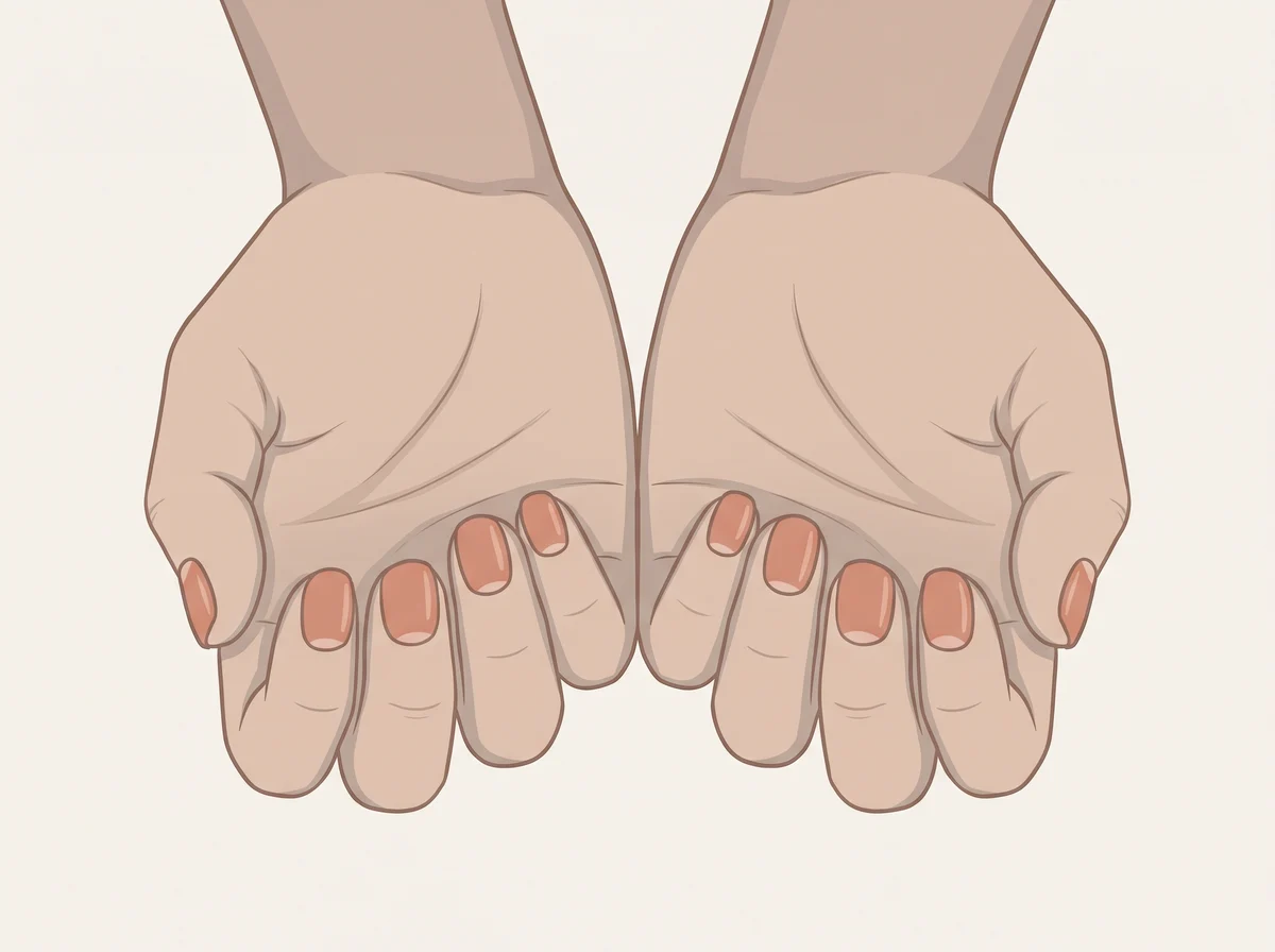
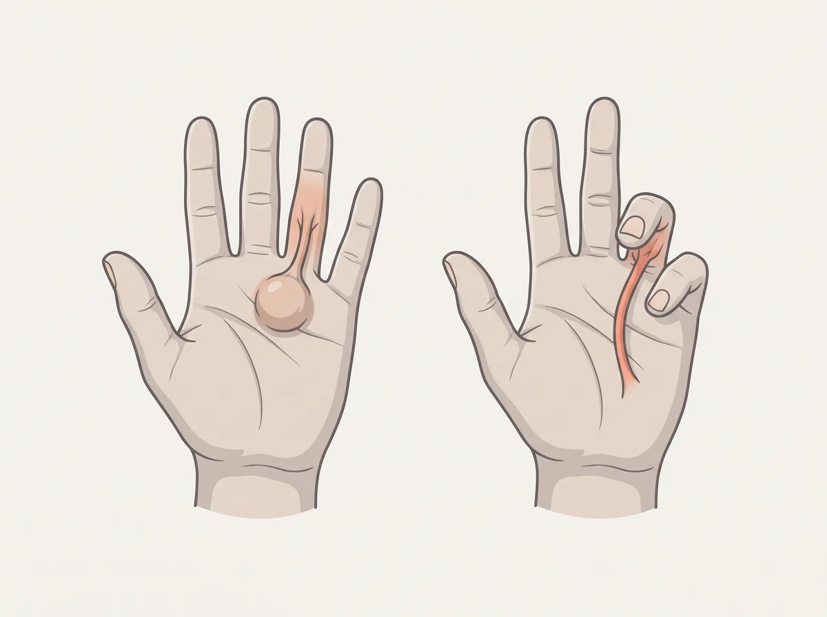
Illustration of a cord in the palm that bends a finger. A firm lump or pit in the palm, usually below the ring or pinky finger, that grows into a tight cord.  Over months or years the cord pulls the finger toward the palm, and you can no longer lay your hand flat on a table.
Illustration, not a photograph
34

A cord in the palm that bends a finger

Dupuytren contracture

A palm cord that curls a finger is worth a blood sugar check

Mention at a checkup

Linked to
  • Dupuytren disease
  • Diabetes
  • Heavy alcohol use
  • Smoking
What to look for
A firm lump or pit in the palm, usually below the ring or pinky finger, that grows into a tight cord. Over months or years the cord pulls the finger toward the palm, and you can no longer lay your hand flat on a table.
Why it happens
The tough tissue layer under the skin of the palm starts to make extra scar-like collagen. Special cells in this tissue tighten and shorten it into a cord that pulls on the finger. Diabetes and heavy alcohol use make this more likely, though no one knows exactly why.
Other causes
Genes are the biggest factor, and it runs in families, mostly in men over 50 with Northern European roots. Epilepsy, past hand injuries, and trigger finger (a finger that catches or locks) can also be involved or look similar.
How strong is the link
Real link A bent finger with a palm cord is a clear sign of Dupuytren disease itself. Studies consistently link it to diabetes, heavy drinking, and smoking, and a 2026 genetic study supports diabetes and alcohol as true causes, but most people with Dupuytren have neither.
What to check
  • Tabletop test: try to lay the palm and fingers flat on a table
  • Hand exam by a doctor or hand surgeon
  • HbA1c or fasting glucose for diabetes
  • Liver function tests if alcohol use is heavy

Get seen soon See a doctor if a finger bends so far that you cannot lay your hand flat or it gets in the way of daily tasks. Treatment works best before the bend gets severe.

  1. Dupuytren Contracture · StatPearls, NCBI Bookshelf, 2023
  2. Dupuytren’s contracture · NHS, 2024
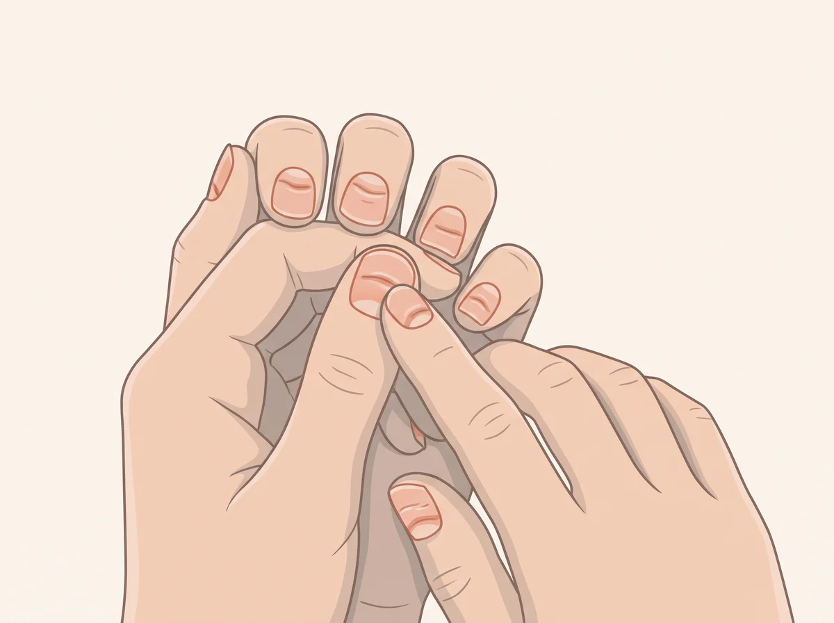
  3. Smoking, alcohol and the risk of Dupuytren’s contracture · Journal of Bone and Joint Surgery, British Volume (Burge et al.), 1997
  4. Causal effects of diabetes and alcohol intake on Dupuytren’s disease: a two-sample Mendelian randomization study in UK Biobank and FinnGen · Korean Journal of Family Medicine (Tian et al.), 2026
Illustration of thin nails that curve up like a spoon. Thin, soft fingernails that dip in the middle and turn up at the edges, like a small spoon.  A drop of water placed on the nail stays in the dip instead of running off.
Illustration, not a photograph
35

Thin nails that curve up like a spoon

Koilonychia

Nails that can hold a drop of water often point to low iron

Mention at a checkup

Linked to
  • Iron deficiency
  • Iron deficiency anemia
  • Hemochromatosis (iron overload)
What to look for
Thin, soft fingernails that dip in the middle and turn up at the edges, like a small spoon. A drop of water placed on the nail stays in the dip instead of running off.
Why it happens
When the body runs low on iron, the nail grows thin and soft. The soft middle sinks while the edges lift into a spoon shape. Too much iron, as in hemochromatosis, can also change how nails grow.
Other causes
Spoon nails are normal in babies and small children, especially on the big toe, and usually go away by about age 9. Frequent contact with oils, solvents, or hair chemicals, as in mechanics and hairdressers, can cause them too.
How strong is the link
Strong link Iron deficiency is the most common cause of spoon nails in adults. The sign is uncommon, showing up in only about 5 percent of people with iron deficiency, so normal nails do not rule out low iron.
What to check
  • Complete blood count (CBC)
  • Ferritin
  • Iron panel (serum iron, transferrin saturation, TIBC)
  • If iron is low without a clear reason, ask about checks for blood loss such as stool tests or a colonoscopy
  • If iron is high, ask about hemochromatosis gene testing

Get seen soon See a doctor soon if spoon nails come with tiredness, shortness of breath, heavy periods, black stools, or weight loss you cannot explain.

  1. Spoon Nails · StatPearls, NCBI Bookshelf, 2023
  2. Digital Clues · American Family Physician (Danoff, Todd), 2018
Illustration of nails that split and peel in layers. The tip of the nail splits into thin, flat layers that flake off, a bit like pastry.  The nail may also feel soft and bendy, or crack at the edge.
Illustration, not a photograph
36

Nails that split and peel in layers

Brittle nail syndrome (onychoschizia)

Nails that peel like pastry often just spend too long in water

Just a quirk

Linked to
  • Lots of water and soap on your hands
  • Normal aging
  • Low iron (less often)
  • Underactive thyroid (less often)
What to look for
The tip of the nail splits into thin, flat layers that flake off, a bit like pastry. The nail may also feel soft and bendy, or crack at the edge.
Why it happens
A nail is made of many thin layers of keratin, a tough protein, stuck together. Getting wet and drying out again and again makes the nail swell and shrink, which loosens those layers at the tip. Harsh soaps, cleaners, and lots of manicures dry the nail out even more.
Other causes
Brittle nails get more common with age, especially in women over 50. Less often, low iron or an underactive thyroid plays a part.
How strong is the link
Real link Dermatology reviews agree that water, soap, and age cause most cases, and up to 1 in 5 people have brittle nails. Biotin pills have only small, uncontrolled studies behind them, and a review found too little proof to use them in healthy people.
What to check
  • Nothing to test for peeling alone. Cut down on time with wet hands first and see if new nail grows in stronger.
  • Ask about a ferritin (iron stores) test if you also feel tired or look pale
  • Ask about a TSH thyroid test if you also feel cold, tired, or gain weight without trying
  1. Optimal diagnosis and management of common nail disorders · Annals of Medicine, 2022
  2. Pathogenesis, Clinical Signs and Treatment Recommendations in Brittle Nails: A Review · Dermatology and Therapy, 2020
  3. Brittle Nails · Canadian Dermatology Association
  4. A Review of the Use of Biotin for Hair Loss · Skin Appendage Disorders, 2017
Illustration of fine lines running from the cuticle to the tip. Thin raised lines run the long way down the nail, from the cuticle to the tip, like the grain in wood.  They are easiest to see when light hits the nail from the side.
Illustration, not a photograph
37

Fine lines running from the cuticle to the tip

Longitudinal ridging (onychorrhexis when marked)

Those fine nail ridges are a lot like wrinkles for your nails

Just a quirk

Linked to
  • Normal aging
  • Dry skin or eczema
  • Underactive thyroid (rarely)
What to look for
Thin raised lines run the long way down the nail, from the cuticle to the tip, like the grain in wood. They are easiest to see when light hits the nail from the side.
Why it happens
Your nail grows from a patch of cells under the cuticle called the matrix. With age, those cells renew more slowly, so the nail grows out with fine grooves. A review of nails in older adults lists these lines as a normal texture change of aging.
Other causes
Very dry skin or eczema can add ridges. An underactive thyroid can cause them too, but then the nails are usually also thick, brittle, or crumbly.
How strong is the link
Strong link In a study of 400 adults, 45.5% of people over 60 had these lines, a much higher share than younger adults. Cleveland Clinic says ridges from aging are not dangerous, so the popular idea that they reveal a hidden deficiency does not hold for fine, even lines.
What to check
  • Nothing to test. Mention it if a single nail changes on its own, such as one deep groove, a split that keeps coming back, or a new dark stripe.

Get seen soon Book a visit soon if one nail gets a new dark brown or black stripe running along it, since that needs a closer look.

  1. Ridges in Nails: Horizontal, Vertical, Causes and Treatment · Cleveland Clinic, 2022
  2. Nail changes and disorders in elderly Egyptians · Journal of Cosmetic Dermatology, 2014
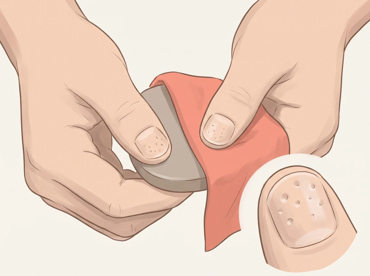
  3. Nails in older adults · Annals of Medicine, 2024
Illustration of no white half-moons at the base of your nails. Look at the base of each nail, just past the cuticle.  Many people see a pale, curved patch there, most often on the thumbs.
Illustration, not a photograph
38

No white half-moons at the base of your nails

Absent lunula (anolunula)

Missing nail half-moons are usually just hiding under your cuticle

Just a quirk

Linked to
  • Normal variation
  • Aging
  • Half-moon hidden under the skin fold
  • Long-term kidney disease (rarely)
What to look for
Look at the base of each nail, just past the cuticle. Many people see a pale, curved patch there, most often on the thumbs. Some people see it on only a few nails, or on none.
Why it happens
The half-moon, or lunula, is the visible front edge of the matrix, the area that makes new nail. Everyone has one, but the skin fold at the base of the nail can cover it. Half-moons are usually biggest on the thumbs and tend to shrink with age.
Other causes
Missing half-moons show up more often in people with long-term kidney disease. Doctors only pay attention to them alongside other symptoms.
How strong is the link
Mostly myth The popular claim that missing half-moons mean a thyroid problem or anemia is a myth. Cleveland Clinic notes that doctors would not diagnose any disease just from looking at the lunulae, and one study saw smaller half-moons in 49% of people over 60.
What to check
  • Nothing to test. Mention it if your half-moons change color, such as turning red, blue, or brown.
  1. What Do the Half-Moons on Your Nails Mean? · Cleveland Clinic, 2025
  2. The lunula · Journal of the American Academy of Dermatology, 1996
  3. Nail changes and disorders in elderly Egyptians · Journal of Cosmetic Dermatology, 2014
  4. Mucocutaneous Manifestations in Patients with Chronic Kidney Disease: A Cross-sectional Study · Indian Dermatology Online Journal, 2018
Illustration of deep sideways grooves across the nails. A groove runs side to side across the nail, and you can feel it with a fingertip.  When something body-wide caused it, the groove sits at about the same spot on most or all nails.
Illustration, not a photograph
39

Deep sideways grooves across the nails

Beau’s lines

A dent across your nails is a little diary entry from a past illness

Just a quirk

Linked to
  • Past severe illness or high fever
  • Chemotherapy
  • Uncontrolled diabetes
  • Zinc deficiency
What to look for
A groove runs side to side across the nail, and you can feel it with a fingertip. When something body-wide caused it, the groove sits at about the same spot on most or all nails.
Why it happens
A big stress on the body, like a high fever, makes the nail root pause for a short time. When growth restarts, the missed stretch shows up as a dent that moves out with the nail. Nails grow about 1 mm every 6 to 10 days, so a groove 5 mm from the cuticle points to an event about one to two months earlier.
Other causes
A bump to one finger, a rough manicure, or extreme cold can make a groove on a single nail. Grooves from a known illness grow out in about six months once you recover. If grooves show up on all your nails and you cannot link them to an illness or new treatment, mention it at your next checkup.
How strong is the link
Strong link Beau’s lines are a well-established sign that something disrupted nail growth, such as fever, chemotherapy, severe malnutrition, or diabetes. The sign tells you that a stress happened and roughly when, but it does not tell you what the stress was.
What to check
  • Look back at illnesses, fevers, surgery, or new medicines one to three months before the groove formed
  • Fasting glucose or HbA1c
  • Complete blood count and basic metabolic panel if there is no clear cause
  • Zinc level if your diet is limited or you have a gut absorption problem
  1. Beau’s lines · BMJ Case Reports, 2018
  2. Beau’s Lines: What It Looks Like, Causes & Treatment · Cleveland Clinic, 2022
  3. Nail Abnormalities: Clues to Systemic Disease · American Family Physician, 2004
Illustration of tiny dents or pits in the nails. Small dents on the nail surface, like the dimples on a thimble.  They can be scattered or sit in rows, and they catch the light when you tilt the nail.
Illustration, not a photograph
40

Tiny dents or pits in the nails

Nail pitting

Tiny thimble dents in your nails are often a psoriasis clue

Mention at a checkup

Linked to
  • Psoriasis
  • Psoriatic arthritis
  • Alopecia areata
What to look for
Small dents on the nail surface, like the dimples on a thimble. They can be scattered or sit in rows, and they catch the light when you tilt the nail.
Why it happens
Psoriasis and alopecia areata are immune conditions that can irritate the nail root. Small spots in the root make faulty nail cells, and those cells flake off the surface as the nail grows. Each missing spot leaves a tiny pit.
Other causes
Less often, pits come with reactive arthritis, sarcoidosis, or the blistering skin disease pemphigus.
How strong is the link
Strong link Pitting affects 10% to 50% of people with psoriasis, and nail changes can be an early clue to psoriatic arthritis. In a 2024 study of 197 people with alopecia areata, pitting was the most common nail change.
What to check
  • Skin check for scaly patches on the scalp, elbows, knees, or belly button
  • Ask about joint pain, morning stiffness, or a swollen finger or toe
  • Dermatology exam of the nails
  • Nail clipping test for fungus if the nail is also thick or crumbly

Get seen soon See a doctor soon if pitted nails come with swollen, stiff, or painful joints. Early treatment of psoriatic arthritis helps protect the joints.

  1. Nail Abnormalities: Clues to Systemic Disease · American Family Physician, 2004
  2. What is nail psoriasis, and how can I treat it? · American Academy of Dermatology, 2023
  3. Clinical and demographic characteristics associated with nail involvement in alopecia areata: A cross-sectional study of 197 patients · Health Science Reports, 2024
Illustration of small white spots on the nails. One or a few small white dots or flecks on a nail.  They appear in different places on different nails and move toward the tip as the nail grows.
Illustration, not a photograph
41

Small white spots on the nails

Punctate leukonychia

White nail spots are usually souvenirs from a bump you forgot

Just a quirk

Linked to
  • Minor nail injury
  • Manicure or nail product damage
  • Fungal nail infection
  • Zinc deficiency (rare)
What to look for
One or a few small white dots or flecks on a nail. They appear in different places on different nails and move toward the tip as the nail grows.
Why it happens
A knock or pressure on the nail root disturbs a few nail cells as they form. Those cells stay slightly cloudy, and they reflect light as a white spot. Pushing back cuticles hard, biting nails, and frequent polish or artificial nails can do the same.
Other causes
A white, chalky patch that scrapes off can be a surface fungal infection. Psoriasis and alopecia areata can also cause white spots, and zinc deficiency is a rare listed cause.
How strong is the link
Mostly myth The popular idea that white spots mean low zinc or calcium has no support. A 2022 dermatology review states the spots are not linked to low calcium or iron in the nail and usually come from injury, and a family medicine review calls scattered spots of no significance.
What to check
  • Usually none; watch whether the spot grows out over a few months
  • Nail clipping test for fungus if the white area is chalky, spreading, or scrapes off
  • Liver and kidney tests only if whole nails turn white
  1. Leukonychia: What Can White Nails Tell Us? · American Journal of Clinical Dermatology, 2022
  2. Nail Abnormalities: Clues to Systemic Disease · American Family Physician, 2004
Illustration of nail beds that stay pale after you press them. With your hand at heart level, press a fingernail until the pink under it turns white.  Hold for about 10 seconds, let go, and count how long the pink takes to come back.
Illustration, not a photograph
42

Nail beds that stay pale after you press them

Slow capillary refill

The 2-second fingertip squeeze doctors use to check your blood flow

Mention at a checkup

Linked to
  • Cold hands
  • Poor blood flow to the hands
  • Dehydration
  • Anemia (if nail beds look pale to begin with)
What to look for
With your hand at heart level, press a fingernail until the pink under it turns white. Hold for about 10 seconds, let go, and count how long the pink takes to come back. Color that takes much longer than 2 to 3 seconds, or nail beds that look pale before you even press, are the sign.
Why it happens
Pressing squeezes blood out of the tiny vessels under the nail, called capillaries. When you let go, blood flows back in and the pink returns. A slow return means less blood is reaching your fingertips right then.
Other causes
A cold room, cold hands, older age, and dim light all make the refill look slower. In a study of 1,000 healthy adults, the typical time was 1.9 seconds, and about 1 in 20 took longer than 3.5 seconds.
How strong is the link
Real link Doctors use this test to check blood flow, mostly in sick or dehydrated patients, but in healthy people it shifts with age, sex, and temperature, and one large study called it unreliable on its own. Pale nail beds do point to anemia, yet in large field studies pallor spotted only about half of people with severe anemia.
What to check
  • Redo the test with warm hands in a warm, bright room
  • Complete blood count (CBC) if your nail beds look pale
  • Ask about your circulation if your fingers often turn white, blue, or numb

Get seen soon Get urgent care if slow color return comes with dizziness, fainting, confusion, or cold, clammy skin.

  1. Capillary nail refill test · MedlinePlus, U.S. National Library of Medicine, 2025
  2. Capillary Refill Time · StatPearls, NCBI Bookshelf, 2023
  3. Impact of patient and environmental factors on capillary refill time in adults · American Journal of Emergency Medicine, 2008
  4. Clinical pallor is useful to detect severe anemia in populations where anemia is prevalent and severe · Journal of Nutrition, 1999

Body

43–45
Illustration of sock marks that stay for a while. Everyone gets sock lines.  Look for deep ones that stay long after the socks come off, often with ankles that look a bit puffy.
Illustration, not a photograph
43

Sock marks that stay for a while

Pitting edema (extra fluid in the lower legs)

Sock lines that linger mean a little fluid has settled in your ankles

Mention at a checkup

Linked to
  • Long sitting or standing, heat, or salty meals
  • Heart or kidney problems
  • Liver disease
  • Vein problems (venous insufficiency)
What to look for
Everyone gets sock lines. Look for deep ones that stay long after the socks come off, often with ankles that look a bit puffy. Press a thumb just above the ankle for a few seconds. If a small dent stays, some extra fluid has settled there.
Why it happens
Fluid seeps out of tiny blood vessels into the skin, and gravity pulls it down to the ankles. Sock elastic presses into the puffy skin and leaves a print that fills back in slowly.
Other causes
Long hours of sitting or standing, hot weather, salty meals, and pregnancy are the usual reasons. Some blood pressure pills, such as amlodipine, can do it too. Heart, kidney, liver, and vein problems are the reasons to get it checked when the swelling is new or stays in both legs day after day.
How strong is the link
Strong link A dent that stays after pressing is a well-established sign of extra fluid. Most of the time the cause is everyday, but the NHS advises seeing a doctor when swelling in both legs does not improve after a few days.
What to check
  • A review of your medicines
  • Blood and urine tests for kidney, liver, and thyroid
  • A heart check if the swelling stays in both legs

Get seen soon Get care the same day if only one leg swells suddenly, or it turns red, warm, or painful. Call emergency services if swelling comes with shortness of breath or chest pain.

  1. Edema: Diagnosis and Management · American Family Physician, 2013
  2. Physiology, Edema · StatPearls, NCBI Bookshelf, 2023
  3. Swollen ankles, feet and legs (oedema) · NHS, 2026
Illustration of hands and feet that are always cold. Your hands and feet feel cold to you or to others, even in a room where most people feel fine, and they take a long time to warm up.
Illustration, not a photograph
44

Hands and feet that are always cold

Cold fingers are often your body guarding its warm core

Mention at a checkup

Linked to
  • Normal response to cold or stress
  • Low iron (anemia)
  • Underactive thyroid
  • Raynaud’s phenomenon
What to look for
Your hands and feet feel cold to you or to others, even in a room where most people feel fine, and they take a long time to warm up.
Why it happens
When you are cold or stressed, the small blood vessels in your hands and feet tighten. Less blood flows to your fingers and toes, and more warmth stays around your heart and other organs. Some people have a stronger version of this response, and it can run in families.
Other causes
Low iron, an underactive thyroid, Raynaud’s phenomenon, narrowed arteries, and some medicines can all cause cold hands and feet. With Raynaud’s, fingers turn white, then blue or purple in the cold, then red as blood returns.
How strong is the link
Real link Cold hands are often a normal, healthy response to cold. About half of people with an underactive thyroid feel the cold more than others, and cold hands and feet are a listed sign of iron-deficiency anemia, so the clue matters most when you also feel tired, short of breath, or run down.
What to check
  • Iron blood tests, including ferritin, to look for low iron
  • Thyroid blood test
  • Mention it if your fingers or toes turn white or blue in the cold

Get seen soon Get care right away if a cold hand or foot changes color and does not improve with warming, or if you get sores on your fingers or toes.

  1. 7 Reasons Why Your Hands and Feet Are Always Cold · Cleveland Clinic Health Essentials, 2026
  2. Iron-Deficiency Anemia · Cleveland Clinic, 2024
  3. Hypothyroidism (Underactive Thyroid) · National Institute of Diabetes and Digestive and Kidney Diseases (NIDDK), 2021
  4. Raynaud’s · NHS
Illustration of a waist bigger than half your height. Wrap a tape around your waist halfway between your lowest rib and the top of your hip bone, which is usually just above the belly button, and breathe out normally.  Divide that number by your height in the same units.
Illustration, not a photograph
45

A waist bigger than half your height

Waist-to-height ratio of 0.5 or more (central adiposity)

The simple tape test that beats BMI at spotting belly-fat risk

Mention at a checkup

Linked to
  • Extra fat around the middle
  • Type 2 diabetes risk
  • High blood pressure
  • Heart disease risk
What to look for
Wrap a tape around your waist halfway between your lowest rib and the top of your hip bone, which is usually just above the belly button, and breathe out normally. Divide that number by your height in the same units. A result from 0.5 to 0.59 means raised risk, and 0.6 or more means higher risk.
Why it happens
A bigger waist usually means more fat stored around the middle of the body. NICE, the UK health guidance body, links this central fat to type 2 diabetes, high blood pressure, and heart disease. Dividing by height lets one simple cutoff work for short and tall people.
Other causes
Pregnancy, bloating, or a tape in the wrong spot can push the number up. NICE says the ratio works for adults with a BMI under 35, including very muscular people.
How strong is the link
Strong link A 2012 review of studies with more than 300,000 adults found waist-to-height ratio picked out diabetes, high blood pressure, and heart risk better than BMI or waist size alone. The 0.5 cutoff held up in studies from 14 countries, and NICE adopted it in 2022.
What to check
  • Blood pressure
  • HbA1c or fasting glucose
  • Cholesterol panel (lipids)
  • Recheck your own ratio every few months
  1. Overweight and obesity management (NG246): Identifying and assessing overweight, obesity and central adiposity (waist-to-height recommendations from 2022) · NICE, 2025
  2. Waist-to-height ratio is a better screening tool than waist circumference and BMI for adult cardiometabolic risk factors: systematic review and meta-analysis · Obesity Reviews, 2012
  3. A systematic review of waist-to-height ratio as a screening tool for the prediction of cardiovascular disease and diabetes: 0.5 could be a suitable global boundary value · Nutrition Research Reviews, 2010

Cravings & quirks

46–50
Illustration of craving and chewing lots of ice. You want to crunch ice every day, often by the cupful or trayful, and the urge is hard to skip.  Liking a cold drink on a hot day does not count.
Illustration, not a photograph
46

Craving and chewing lots of ice

Pagophagia (a type of pica)

An ice-chewing habit can be your body waving a low-iron flag

Mention at a checkup

Linked to
  • Iron deficiency
  • Iron-deficiency anemia
  • Pregnancy
What to look for
You want to crunch ice every day, often by the cupful or trayful, and the urge is hard to skip. Liking a cold drink on a hot day does not count.
Why it happens
Nobody knows the exact reason. In one small study, people with anemia got faster and sharper on a test after chewing ice, while healthy people did not change, possibly because chewing ice sends more blood to the brain. The craving often fades within days to weeks once iron is replaced, sometimes before blood counts recover.
Other causes
Ice chewing can also come from habit, stress, or culture. It is common in children and during pregnancy.
How strong is the link
Strong link In a clinic study of 262 adults with low iron, 45% had pica, and 87% of those craved ice. In another study, only 4% of people without anemia chewed ice this way, compared with 56% of people with anemia.
What to check
  • Ferritin (iron stores)
  • Complete blood count (CBC)
  • Ask what might be draining your iron, such as heavy periods
  1. Pica associated with iron deficiency or depletion: clinical and laboratory correlates in 262 non-pregnant adult outpatients · BMC Blood Disorders, 2010
  2. Pagophagia improves neuropsychological processing speed in iron-deficiency anemia · Medical Hypotheses, 2014
  3. Pagophagia in iron deficiency anemia · Rinsho Ketsueki, 2014
  4. Craving for ice and iron-deficiency anemia: a case series from Oman · Pediatric Hematology and Oncology, 2005
Illustration of strong salt cravings. You keep reaching for chips, pickles, or the salt shaker, or food tastes flat until it is very salty.  Notice when it happens, such as after exercise, on a hot day, or before your period.
Illustration, not a photograph
47

Strong salt cravings

Salt cravings after a sweaty workout are your body doing its job

Mention at a checkup

Linked to
  • Heavy sweating
  • Stress, poor sleep, or period hormones
  • POTS (a racing heart when you stand)
  • Addison disease (rare)
What to look for
You keep reaching for chips, pickles, or the salt shaker, or food tastes flat until it is very salty. Notice when it happens, such as after exercise, on a hot day, or before your period.
Why it happens
Sweat carries salt out of your body, so after heavy sweating your body nudges you to replace it. Stress hormones, poor sleep, and hormone shifts before a period can also stir up salty cravings. In Addison disease, the adrenal glands make too little of the hormones that help you hold on to salt, so the body keeps losing it.
Other causes
Boredom and habit play a big part too. Doctors often tell people with the low-blood-volume type of POTS to eat more salt and drink more fluid.
How strong is the link
Real link Salt craving is a textbook symptom of Addison disease, which is rare. Everyday causes like sweating, stress, and habit are far more common, though few studies measure how often a craving points to illness.
What to check
  • Blood sodium and potassium (basic metabolic panel)
  • Ask about a cortisol blood test if cravings come with tiredness, weight loss, or darker patches of skin
  • Ask about POTS if your heart races and you feel faint when you stand up

Get seen soon Book a visit soon if salt cravings come with weight loss, dizziness when you stand, and skin that looks darker than usual.

  1. Why Do You Crave Salt? · Cleveland Clinic, 2020
  2. Addison’s disease: Symptoms · NHS, 2025
  3. Addison’s Disease · Cleveland Clinic, 2026
  4. Postural Orthostatic Tachycardia Syndrome (POTS) · Cleveland Clinic, 2026
Illustration of pink or red pee after eating beets. Your pee turns pink, red, or purplish after you eat beets, drink beet juice, or eat foods colored with beet.  It fades as the beets pass through your system.
Illustration, not a photograph
48

Pink or red pee after eating beets

Beeturia

Pink pee after borscht is just beet pigment passing through

Just a quirk

Linked to
  • Beet pigment passing into your pee
  • Iron deficiency
  • Gut absorption problems (rare)
What to look for
Your pee turns pink, red, or purplish after you eat beets, drink beet juice, or eat foods colored with beet. It fades as the beets pass through your system.
Why it happens
Beets get their red color from pigments called betacyanins. In most people little of that pigment reaches the urine, but some people absorb and pass enough to tint it. Scientists once blamed food allergies or bladder bacteria, but later studies ruled those ideas out.
Other causes
Red or brown pee when you have not eaten beets can mean blood in the urine or a side effect of a medicine. That kind needs a check.
How strong is the link
Real link StatPearls puts beeturia at 10% to 14% of people and says it is more common with iron deficiency. The iron link comes from older, small studies, so pink pee alone cannot diagnose low iron.
What to check
  • Nothing to test for beeturia itself. If you also feel tired or look pale, ask for a ferritin (iron stores) test.

Get seen soon See a doctor soon if your pee is red or brown and you have not eaten beets, or if the color does not clear within a couple of days.

  1. Beeturia · StatPearls, NCBI Bookshelf, 2023
  2. Food idiosyncrasies: beetroot and asparagus · Drug Metabolism and Disposition, 2001
  3. Beeturia. A sign of iron deficiency · American Journal of Diseases of Children, 1969
Illustration of pee that smells after asparagus, or a nose that cannot smell it. Your pee has a strong sulfur or cooked-cabbage smell soon after you eat asparagus.  Or you have eaten plenty of asparagus and never noticed a thing.
Illustration, not a photograph
49

Pee that smells after asparagus, or a nose that cannot smell it

Asparagus anosmia (for people who cannot smell it)

Asparagus pee is real, and about 6 in 10 people cannot smell it

Just a quirk

Linked to
  • Sulfur compounds from asparagus
  • Your smell genes
  • Nothing to worry about
What to look for
Your pee has a strong sulfur or cooked-cabbage smell soon after you eat asparagus. Or you have eaten plenty of asparagus and never noticed a thing.
Why it happens
Your body breaks down compounds in asparagus into sulfur chemicals, such as methanethiol, that leave in your pee. A lab study found that people differ both in how much of the smelly chemicals they make and in whether they can smell them. A large genetic study tied the ability to smell it to a cluster of smell-receptor genes.
Other causes
A strong, unpleasant pee smell that shows up without asparagus has other causes. If it comes with burning when you pee, get it checked.
How strong is the link
Strong link A 2016 BMJ study of 6,909 adults found that 58% of men and 61.5% of women could not smell asparagus pee, and linked this to 871 genetic variants near smell genes. A smaller lab study confirmed that both making and smelling the odor vary between people.
What to check
  • Nothing to test. It makes a fun dinner-table experiment.
  1. Sniffing out significant “Pee values”: genome wide association study of asparagus anosmia · BMJ, 2016
  2. Excretion and perception of a characteristic odor in urine after asparagus ingestion: a psychophysical and genetic study · Chemical Senses, 2011
  3. Food idiosyncrasies: beetroot and asparagus · Drug Metabolism and Disposition, 2001
Illustration of sneezing when you step into bright sun. One or more sneezes right after you walk from a dim place into bright sunlight.  It happens again and again in the same way.
Illustration, not a photograph
50

Sneezing when you step into bright sun

Photic sneeze reflex (ACHOO syndrome)

About 1 in 4 people sneeze when they step into the sun

Just a quirk

Linked to
  • Inherited genes
  • A harmless crossed wire between light and sneezing
What to look for
One or more sneezes right after you walk from a dim place into bright sunlight. It happens again and again in the same way.
Why it happens
Nobody knows the full answer. One idea is that the trigeminal nerve, which senses touch in the face and nose, gets pulled in when the eyes see a burst of light. Another is that the brain’s vision area reacts extra strongly to light in sun sneezers.
Other causes
It is a harmless trait. The playful name ACHOO stands for Autosomal Dominant Compulsive Helio-Ophthalmic Outburst, and it often runs in families.
How strong is the link
Real link In a 2019 study of 3,417 people in China, 25.6% said they sneeze in bright light, and gene scans found several regions tied to it. Rates vary by country, and nobody has pinned down how it works.
What to check
  • Nothing to test.
  • Sunglasses or a hat can head off the sneeze, which helps when you drive out of a tunnel.
  1. ACHOO Syndrome · Medical Genetics Summaries, NCBI Bookshelf, 2015
  2. A genome-wide association study on photic sneeze reflex in the Chinese population · Scientific Reports, 2019

What to do with this

Count the ones you spotted. Most people find a few harmless quirks. If several point to the same thing, write down when each one started and bring the list to your next checkup. A simple blood panel covers most of this guide: complete blood count, ferritin, B12, vitamin D, TSH with free T4, a lipid panel, and HbA1c.

This guide is education, not medical advice, and it cannot diagnose you. Every picture in it is an illustration drawn for this guide, not a photograph of a real person. Each one shows a typical version of the sign, and yours will look different. Use them to spot something worth asking about, never to rule something out. If something is sudden, painful, or spreading, get it checked the same day.

Spotted something familiar?

Save the guide so you can note the changes and bring your questions to your next checkup.

Look inside first

Three real pages, before you give an email.

No sign-up to see these. They are unedited pages from the 53-page PDF, so you can judge the guide instead of a cover.

Page 2 of 50 Body Signals: All 50, on one spread
PAGE 2

All 50, on one spread

Every signal with the thing it points to, so you can scan for the one you came here about.

Page 9 of 50 Body Signals: One signal, fully worked
PAGE 9

One signal, fully worked

What to look for, why it happens, what else causes it, how strong the link is, and what to ask for.

Page 50 of 50 Body Signals: Most of them are harmless
PAGE 50

Most of them are harmless

Plenty of entries end with nothing to test. The guide is as clear about those as it is about the rest.

Honest fit

Who this helps, and who should skip it.

Worth your time if

  • You noticed something change and want to know whether it means anything.
  • You want to walk into a checkup with a specific question instead of a vague worry.
  • You want to see what each change looks like, not just read a description of it.

Skip it if

  • You want a diagnosis. The illustrations show a typical version and yours will look different.
  • Something is sudden, painful or spreading. That needs care today, not a PDF.

Before you download

The questions people actually ask.

Is this going to make me anxious about nothing?

Most of these are harmless, and the guide says so in the first rule on page 2. Each signal carries a seriousness tag and an evidence rating, so a weak link looks weak. The useful pattern is two or three signals pointing the same way, which is the thing the guide is built to help you notice.

Are the pictures real photos?

No. Every one is an illustration drawn for this guide, and each is labelled as one. They are there to show you what a change looks like, and they are deliberately not photographs of a real person. Use them to recognise something worth asking about, never to rule something out.

Can I just read the page instead of downloading?

Yes. All 50 signals are on this page in full, free and ungated. The PDF is for reading offline, printing, or taking to an appointment.

Why is it free?

Because I want more people running careful experiments on themselves, and because some readers eventually become Rebody Health customers. That is the whole model. No upsell arrives with the guide.

What happens to my email address?

You get the guide, then AI Biohacking, which is what I am testing and measuring on myself. You also get the revised guide whenever the research changes. One click unsubscribes and the address is never sold or shared.

What you are getting

1,000+ people already have this guide.

  • 50 signals with photos
  • Evidence rated for every one
  • Seriousness flagged
  • What to check, per signal
Last checked
September 27, 2026. I recheck the sources behind each signal and the license on every photo, and revise the guide when the evidence changes.
When it changes
The revised guide goes to everyone on the list. You do not have to come back for it.
Who wrote it
Nick Locascio. AI entrepreneur, founder of Rebody Health, co-founder of Booth AI (YC). My health experiments are personal experience, not medical advice. See the work behind it.

The rest of the field guides

Every one is free, and built the same way.

© 2026 Nick Locascio

Personal experiments and research. Educational information.